Structure of Oral Tissues: A Complete Beginner’s Guide
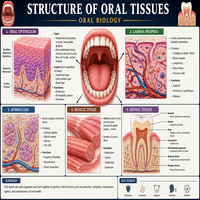
The mouth contains many specialized tissues. Each tissue plays a unique role in chewing, speaking, and protecting deeper structures. Therefore, dental students must understand the structure of oral tissues before moving into clinical subjects.
This guide explains every major oral tissue in a simple, organized way. Furthermore, it includes tables and a flowchart to make revision easier. As a result, even complete beginners can follow along without confusion.
What Is the Structure of Oral Tissues?
The structure of oral tissues includes both hard and soft components. Hard tissues, for instance, include enamel, dentin, cementum, and bone. Soft tissues, however, include oral mucosa, salivary glands, and the periodontal ligament.
Below is a simple flowchart that shows this classification clearly.
                    ORAL TISSUES
                         |
        -------------------------------------
        |                                   |
   Hard Tissues                       Soft Tissues
        |                                   |
 -------------------                -------------------
 |    |     |       |               |        |         |
Enamel Dentin Cementum Bone     Oral Mucosa  Salivary  Periodontal
                                              Glands    Ligament
Consequently, this classification helps students group similar structures together. Once you understand this division, individual tissues become easier to learn in detail.
The Tooth: Hard Tissue Composition
The tooth forms the most mineralized structure in the human body. Specifically, it consists of four main tissues, each with a distinct function.
| Tissue | Composition | Function |
|---|---|---|
| Enamel | 96% mineral, 4% organic matter | Protects the crown from wear |
| Dentin | 70% mineral, 30% organic matter | Supports enamel, transmits sensation |
| Cementum | 65% mineral, 35% organic matter | Anchors periodontal fibers to the root |
| Pulp | Soft connective tissue | Supplies nerves and blood vessels |
Notably, enamel contains no living cells once it forms. Dentin and cementum, on the other hand, remain biologically active throughout life. Hence, these tissues can respond to certain stimuli, such as decay or wear.
Supporting Tissues of Tooth
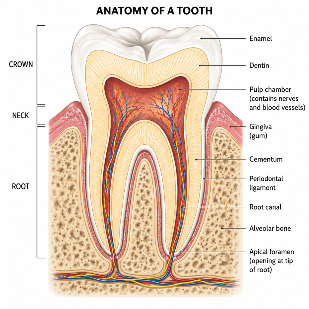
The supporting tissues of tooth, often called the periodontium, hold the tooth firmly within the jaw. Without these tissues, teeth would lack stability during chewing.
| Component | Description |
|---|---|
| Gingiva | Soft tissue surrounding the tooth neck |
| Periodontal ligament | Fibrous tissue connecting tooth root to bone |
| Cementum | Hard layer covering the root surface |
| Alveolar bone | Bone socket that houses the tooth root |
The periodontal ligament absorbs chewing forces and protects the bone from damage. Meanwhile, the gingiva forms a protective seal around the tooth. Together, these supporting tissues of tooth maintain function and prevent infection from reaching deeper structures.
Oral Mucosa
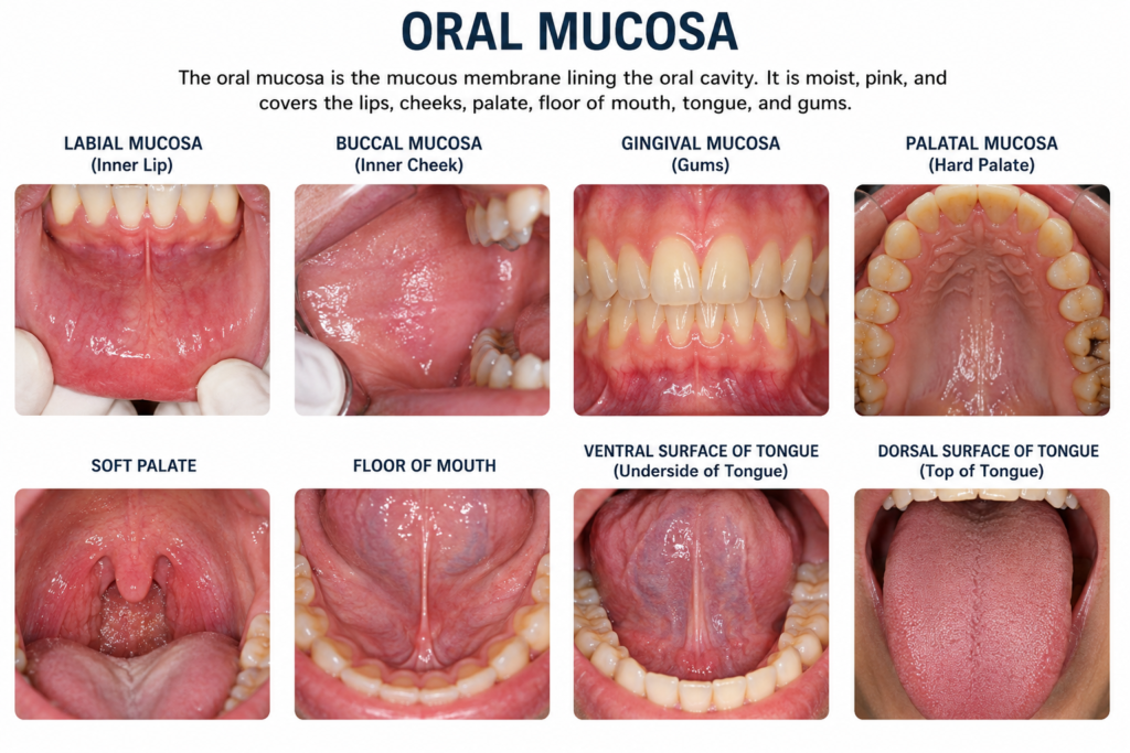
Oral mucosa lines the entire inside of the mouth. It differs in thickness and texture depending on its location and function.
| Type | Location | Feature |
|---|---|---|
| Masticatory mucosa | Gingiva, hard palate | Keratinized, tough surface |
| Lining mucosa | Cheeks, lips, soft palate | Non-keratinized, flexible |
| Specialized mucosa | Dorsum of tongue | Contains taste buds |
Masticatory mucosa resists friction during chewing. Lining mucosa, in contrast, allows movement of the cheeks and lips. Similarly, specialized mucosa supports sensory functions, such as taste perception. Overall, this variation reflects the diverse roles oral mucosa performs daily.
Salivary Glands
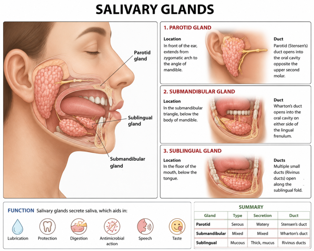
Salivary glands produce saliva, which aids digestion, lubrication, and protection against bacteria. Three major pairs of glands contribute most of the saliva produced each day.
| Gland | Location | Secretion Type |
|---|---|---|
| Parotid gland | In front of the ear | Serous (watery) |
| Submandibular gland | Below the jaw | Mixed (serous and mucous) |
| Sublingual gland | Under the tongue | Mainly mucous |
Additionally, many small minor salivary glands exist throughout the oral cavity. These glands keep the mucosa moist between meals. Consequently, reduced saliva flow can lead to dryness, discomfort, and a higher risk of decay.
Bones of Jaw and the TMJ
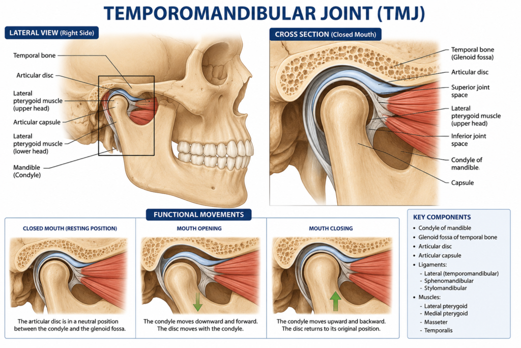
The bones of jaw form the framework that supports teeth and facial structures. Two main bones make up this framework: the maxilla and the mandible.
| Bone | Type | Key Feature |
|---|---|---|
| Maxilla | Fixed bone | Forms the upper jaw and houses upper teeth |
| Mandible | Movable bone | Forms the lower jaw, allows chewing motion |
The temporomandibular joint, commonly called the TMJ, connects the mandible to the skull. Therefore, this joint allows opening, closing, and side-to-side jaw movement. Without a healthy TMJ, chewing and speaking would become difficult. Similarly, any dysfunction in this joint often causes pain near the ear or jaw.
Hard Tissue Formation and Mineralization
Hard tissue formation begins early during tooth development. Specialized cells, such as ameloblasts and odontoblasts, deposit organic matrix first. Afterward, mineral crystals fill this matrix during a process called mineralization.
| Stage | Process | Cells Involved |
|---|---|---|
| Matrix formation | Organic framework laid down | Ameloblasts, odontoblasts |
| Mineralization | Calcium and phosphate crystals deposited | Same cells, plus enzymes |
| Maturation | Tissue hardens fully | Continued mineral exchange |
Mineralization strengthens enamel and dentin significantly. Indeed, without proper mineralization, teeth would remain soft and prone to damage. Thus, nutrition and fluoride exposure during development directly influence this process.
Hard Tissue Degradation
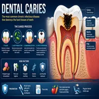
Hard tissue degradation occurs when acids or enzymes break down mineral content faster than the body can repair it. Dental caries represents the most common example of this process.
Bacteria in plaque produce acids after consuming sugars. These acids dissolve mineral crystals within enamel and dentin. Subsequently, if the process continues unchecked, cavities form and progress toward the pulp.
Fortunately, early degradation can reverse through remineralization. Fluoride, saliva, and proper oral hygiene support this natural repair process. Therefore, regular dental visits remain essential for catching damage early.
Quick Summary Table
| Component | Category | Main Role |
|---|---|---|
| Tooth | Hard tissue | Chewing and biting |
| Supporting tissues of tooth | Mixed | Stability and attachment |
| Oral mucosa | Soft tissue | Protection and sensation |
| Salivary glands | Soft tissue | Saliva production |
| Bones of jaw | Hard tissue | Structural framework |
| TMJ | Joint | Jaw movement |
This table offers a quick overview of the structure of oral tissues discussed throughout this guide. Beginners can use it for fast revision before exams.
Conclusion
The structure of oral tissues includes a wide range of hard and soft components, each working together smoothly. Teeth, supporting tissues, oral mucosa, salivary glands, and jaw bones all contribute to proper oral function.
Moreover, understanding mineralization and degradation helps explain how teeth stay strong or become damaged over time. Once these basics feel familiar, more advanced dental topics become much easier to grasp. Therefore, regular revision of these foundational tissues benefits every dental student significantly.
Frequently Asked Questions
It includes teeth, supporting tissues of tooth, oral mucosa, salivary glands, and bones of jaw. Together, these components support chewing, speech, and
They are collectively called the periodontium. This includes gingiva, periodontal ligament, cementum, and alveolar bone.
Mineralization deposits calcium and phosphate crystals into the organic matrix of enamel and dentin. As a result, the tissue becomes hard and resistant to wear.
Bacterial acids break down mineral content faster than the body can repair it. Consequently, this process leads to dental caries if left untreated.
The TMJ connects the jaw to the skull and allows movement needed for chewing and speaking. Therefore, any dysfunction here can affect daily oral function.