The human face hides many small but powerful structures. One of the most important is the parotid region. This area sits just in front of the ear and plays a major role in digestion, facial movement, and even certain diseases. Therefore, understanding its anatomy matters for students, dentists, surgeons, and curious readers alike.
This guide breaks down the parotid region in simple terms. It covers boundaries, contents, blood supply, nerve pathways, and common clinical conditions. Along the way, you will find tables and a flowchart to make the concepts easier to remember.
What Is the Parotid Region?
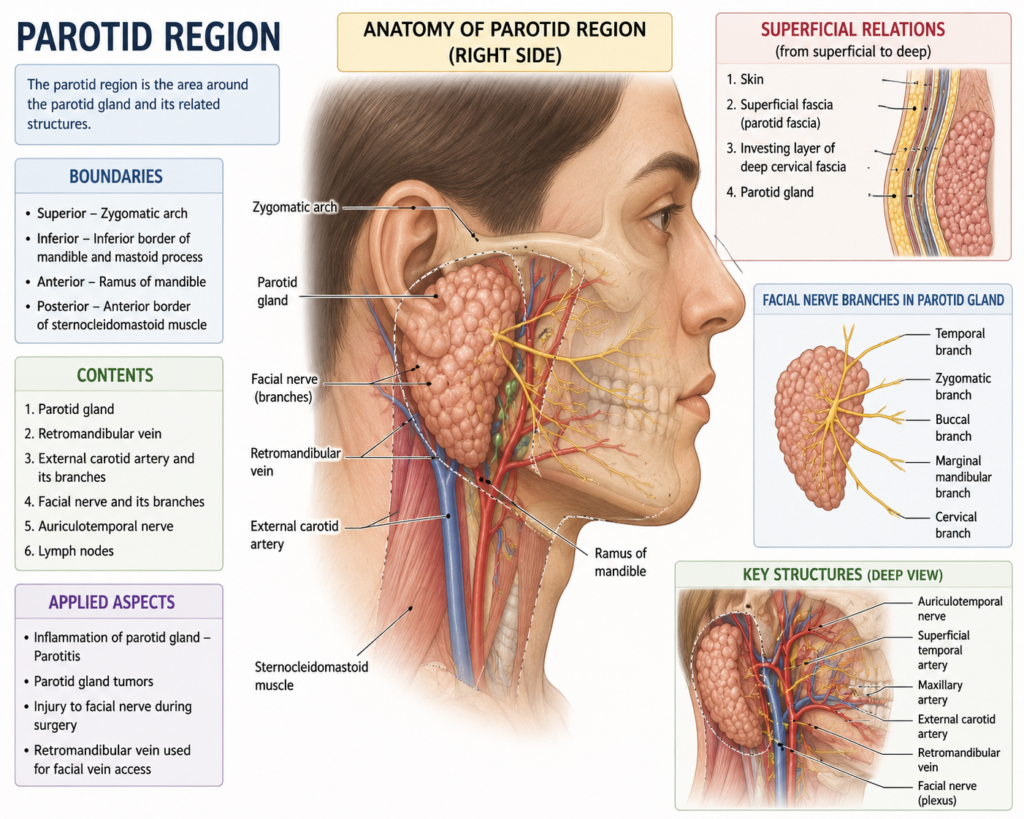
The parotid region refers to the anatomical space located below and in front of the ear, extending toward the angle of the jaw. It houses the largest of the three major salivary glands, the parotid gland. Consequently, this region holds huge clinical importance in dentistry and ENT surgery.
Unlike other salivary glands, the parotid gland does not sit in a simple pouch. Instead, it wraps around several vital structures. As a result, surgeries in this area demand extreme precision. A single wrong cut can damage a nerve responsible for facial expressions.
Interestingly, the gland’s shape resembles an inverted pyramid. Its base touches the skin, while its apex reaches deep near the styloid process. Because of this complex shape, anatomists divide the gland into superficial and deep lobes. The facial nerve runs between these two lobes, acting as a natural dividing line.
In short, the parotid region is not just a single gland. It is a compact zone packed with nerves, vessels, and connective tissue, all working together.
Boundaries of the Parotid Region
Every anatomical region needs clear borders. Otherwise, identifying structures during surgery becomes confusing. The table below summarizes the standard boundaries.
| Boundary | Structure |
|---|---|
| Superior | External auditory meatus and posterior part of the zygomatic arch |
| Inferior | Angle of the mandible |
| Anterior | Masseter muscle |
| Posterior | Mastoid process and sternocleidomastoid muscle |
| Medial | Styloid process and its attached muscles |
| Lateral | Skin and superficial fascia |
These boundaries create a snug compartment. Additionally, a tough fibrous layer called the parotid fascia wraps around the gland. This fascia limits swelling, which explains why infections in this area often cause sharp, throbbing pain.
Furthermore, the fascia connects to deeper neck structures. Because of this connection, infections can sometimes spread beyond the local area if left untreated. For this reason, doctors monitor swelling in this zone carefully.
Contents of the Parotid Region
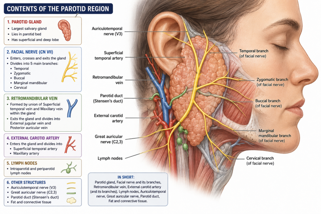
Several structures pass through or lie within this compact space. Below is a simple breakdown.
- Parotid gland – the main salivary gland, divided into superficial and deep lobes
- Facial nerve (CN VII) – controls muscles of facial expression
- Retromandibular vein – formed within the gland by the union of superficial temporal and maxillary veins
- External carotid artery – passes through the gland and divides into terminal branches
- Auriculotemporal nerve – carries sensory and parasympathetic fibers
- Parotid lymph nodes – filter lymph from the scalp, face, and ear
Notably, the facial nerve exits the skull through the stylomastoid foramen. It then enters the gland and splits into five main branches. These branches resemble the fingers of an open hand, spreading toward the forehead, eyes, cheeks, mouth, and neck.
Meanwhile, the external carotid artery enters the gland’s lower part. It travels upward and splits into the maxillary and superficial temporal arteries near the neck of the mandible. This arrangement explains why parotid surgery carries a real risk of bleeding.
Nerve and Blood Supply Flowchart
Visualizing pathways often helps more than reading long paragraphs. Here is a simplified flow of the nerve supply through the parotid region.
Facial Nerve (CN VII)
        |
Exits Stylomastoid Foramen
        |
Enters Parotid Gland
        |
   Splits into 5 Branches
        |
 ---------------------------------
 |     |      |       |          |
Temporal Zygomatic Buccal Marginal Mandibular Cervical
Similarly, the blood supply follows a clear path:
External Carotid Artery
        |
Enters Parotid Gland (posterior border)
        |
Divides Near Neck of Mandible
        |
   ------------------------
   |                      |
Maxillary Artery   Superficial Temporal Artery
These flowcharts show why surgeons treat this region with caution. A small mistake near the branching points can cause permanent facial weakness or heavy bleeding.
Clinical Significance of the Parotid Region
This region is not just an anatomy topic for textbooks. It carries real-world weight in medicine. For instance, mumps, a viral infection, commonly swells the parotid gland and causes visible cheek puffiness.
Similarly, tumors often develop here. Most parotid tumors are benign, such as pleomorphic adenomas. However, some can turn malignant, so early diagnosis stays essential. Doctors typically use ultrasound, CT scans, or fine-needle aspiration to check these growths.
Moreover, surgeons must map the facial nerve carefully before removing any tumor. Otherwise, patients risk losing control over parts of their face. Modern techniques, including nerve monitoring devices, now reduce this risk significantly.
Additionally, dentists sometimes encounter parotid duct stones, known as sialoliths. These stones block saliva flow and cause painful swelling during meals. Warm compresses, hydration, and sometimes minor surgery help resolve this issue.
Common Conditions Affecting the Parotid Region
Below is a quick reference table of conditions linked to this area.
| Condition | Cause | Common Symptom |
|---|---|---|
| Mumps | Viral infection | Painful facial swelling |
| Sialolithiasis | Salivary stone | Pain while eating |
| Pleomorphic adenoma | Benign tumor | Slow-growing painless lump |
| Parotitis | Bacterial infection | Fever, redness, tenderness |
| Bell’s palsy (related nerve) | Facial nerve inflammation | Sudden facial weakness |
Understanding these conditions helps patients seek timely treatment. Early action often prevents complications and speeds up recovery.
Conclusion
The parotid region may look small, yet it holds enormous anatomical and clinical value. It houses a major salivary gland, a critical facial nerve, and important blood vessels, all packed into one compact space. Because of this complexity, both students and clinicians must study it with care.
From boundaries to blood supply, every detail in this region connects to real medical outcomes. Whether you are preparing for an exam or trying to understand a swelling near your jaw, knowing this anatomy gives you a clearer picture. Ultimately, a solid grasp of the parotid region supports safer surgeries, faster diagnoses, and better patient care.
Frequently Asked Questions
1. Where exactly is the parotid region located? It sits just below and in front of the ear, extending toward the angle of the jaw.
2. Why is the facial nerve important in this region? The facial nerve passes directly through the gland and controls all facial expression muscles. Damage during surgery can cause lasting weakness.
3. What causes swelling in the parotid region? Common causes include mumps, bacterial infections, blocked salivary ducts, and tumors.
4. Are parotid tumors always cancerous? No, most parotid tumors are benign. However, doctors still recommend evaluation to rule out malignancy.
5. How do doctors diagnose parotid gland problems? Doctors typically use physical examination, ultrasound, CT scans, or fine-needle aspiration biopsy for accurate diagnosis.
It sits just below and in front of the ear, extending toward the angle of the jaw.
The facial nerve passes directly through the gland and controls all facial expression muscles. Damage during surgery can cause lasting weakness.
Common causes include mumps, bacterial infections, blocked salivary ducts, and tumors.
No, most parotid tumors are benign. However, doctors still recommend evaluation to rule out malignancy.
Doctors typically use physical examination, ultrasound, CT scans, or fine-needle aspiration biopsy for accurate diagnosis.