Tooth Development: Stages, Structure & Supporting Tissues
Every smile hides a fascinating biological story. Long before a baby ever bites into food, tiny cell clusters inside the jaw start working on a lifelong project. This process, called odontogenesis, shapes the enamel, dentin, and roots we rely on every day. It also builds the surrounding structures that hold everything firmly in place.
This guide breaks the entire journey into simple stages. You will see how a single tooth develops, and how its supporting tissues grow alongside it. Whether you are a dental student or simply curious about oral biology, this article keeps things clear and practical.
What Triggers Tooth Development?
Development begins around the sixth week of embryonic life. At this point, the oral epithelium thickens and forms a horseshoe-shaped band called the dental lamina. This band runs along both the upper and lower jaws.
Next, specific spots along the dental lamina start budding inward. These buds mark the exact locations where individual teeth will eventually appear. Meanwhile, the underlying mesenchyme (connective tissue) begins condensing beneath each bud. This interaction between epithelium and mesenchyme drives the entire process forward.
Consequently, dentists often describe tooth formation as a “tissue conversation.” The epithelium signals the mesenchyme, and the mesenchyme signals back. As a result, both tissues shape each other continuously until the structure is complete.
Stage One: The Bud Stage
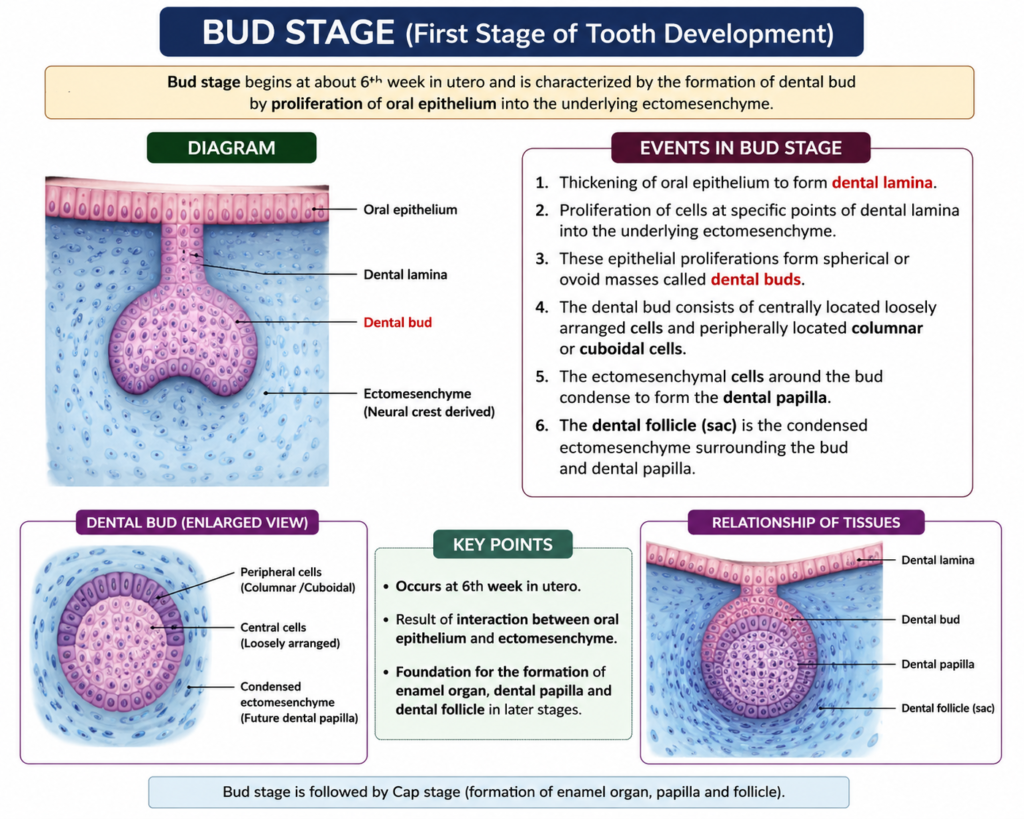
The bud stage kicks off active growth. Each epithelial bud pushes into the mesenchyme, forming a rounded mass of cells. At the same time, the surrounding mesenchymal cells cluster tightly around this bud.
This stage sets the foundation for everything that follows. The bud determines the future size and general position of the crown. Additionally, the number of buds formed corresponds directly to the number of teeth a person will eventually have, including both primary and permanent sets.
Genetics play a huge role here. Therefore, any disruption during this phase can lead to missing teeth (hypodontia) or extra teeth (hyperdontia) later in life.
Stage Two: The Cap Stage
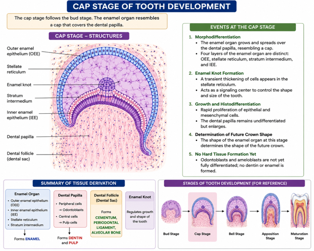
As growth continues, the bud reshapes into a cap-like structure. This happens because cell proliferation is uneven; cells on one side grow faster than others. Consequently, the tissue folds inward, creating the characteristic cap shape.
Four important structures emerge during this stage:
- Enamel organ – forms from the epithelial cap
- Dental papilla – sits inside the cap, made of condensed mesenchyme
- Dental sac (follicle) – surrounds the entire structure
- Stellate reticulum – star-shaped cells inside the enamel organ that support later enamel formation
Together, these three components—the enamel organ, dental papilla, and dental sac—are known as the tooth germ. This germ eventually forms both the visible crown and its supporting tissues.
Stage Three: The Bell Stage
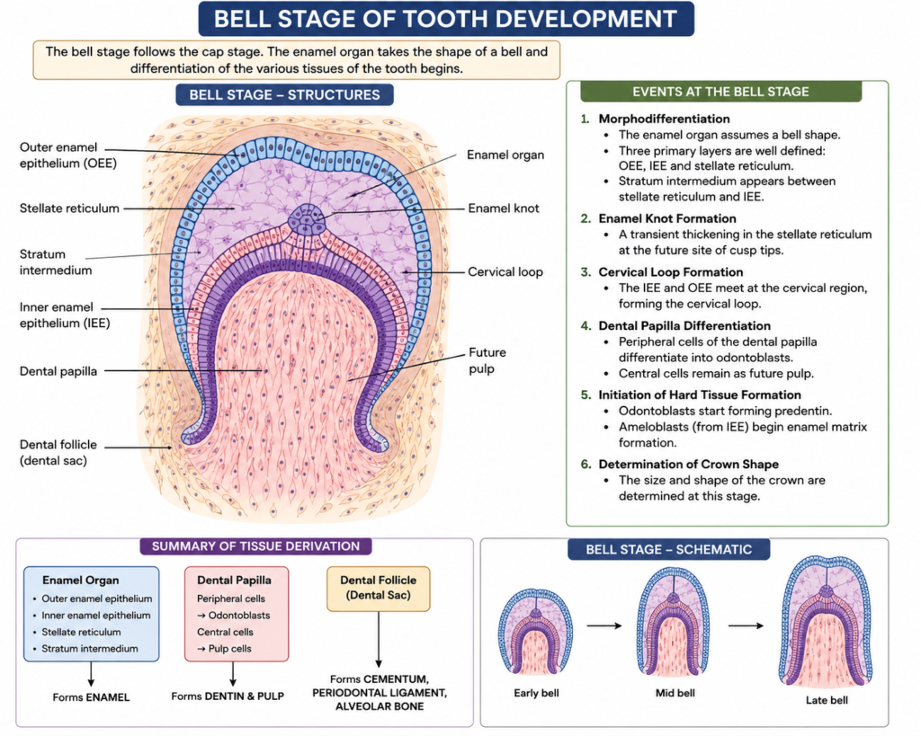
The bell stage brings even more definition. The enamel organ deepens further, taking on a bell shape. Meanwhile, cells begin differentiating into specialized types.
Ameloblasts form along the inner enamel epithelium. These cells later produce enamel. Simultaneously, odontoblasts differentiate from the dental papilla and start producing dentin. This dual differentiation happens almost like a coordinated dance, with signals passing back and forth between the two cell layers.
Furthermore, the shape of the crown becomes fully established during this stage. This determines whether a molar, premolar, incisor, or canine will form at that specific location. Once the crown outline is set, growth shifts toward mineralization and root formation.
Root Formation and the Dentin-Pulp Complex
After crown formation nears completion, Hertwig’s epithelial root sheath takes over. This structure guides root shape and length. It grows downward from the cervical loop, wrapping around the dental papilla.
As the root sheath elongates, odontoblasts continue laying down dentin along the root. Meanwhile, the innermost portion of the dental papilla transforms into dental pulp. This pulp houses nerves and blood vessels, keeping the structure alive and sensitive to stimuli.
Interestingly, root development often continues even after the crown erupts into the mouth. In fact, complete root formation can take two to three years after a tooth becomes visible.
Development of Supporting Tissues
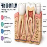
A tooth cannot function alone. It needs strong support from surrounding structures, collectively called the periodontium. This system includes four main components.
| Supporting Tissue | Origin | Primary Function |
|---|---|---|
| Cementum | Dental sac (follicle) | Anchors fibers to the root surface |
| Periodontal ligament (PDL) | Dental sac (follicle) | Absorbs chewing forces, connects root to bone |
| Alveolar bone | Dental sac and surrounding mesenchyme | Houses the root socket |
| Gingiva | Oral ectoderm and mesenchyme | Protects underlying structures, seals the area |
Cementum forms first, laid down by cementoblasts that differentiate from the dental sac. Meanwhile, fibroblasts within the same sac produce collagen fibers that eventually become the periodontal ligament. These fibers connect cementum to the surrounding alveolar bone, creating a flexible yet stable attachment.
Alveolar bone develops through intramembranous ossification. Osteoblasts deposit bone matrix around the developing root, gradually forming the socket. Meanwhile, gingival tissue develops from both oral epithelium and connective tissue, creating a protective seal around the neck of the structure.
This entire support system works together like a shock-absorbing suspension. Without it, chewing forces would damage the root and surrounding bone quickly.
Tooth Eruption Process
Eruption is not simply “pushing through.” It involves active biological remodeling. Bone resorption occurs above the developing structure, creating a pathway toward the oral cavity. Simultaneously, the periodontal ligament fibers reorganize, generating the tension needed for movement.
Here is a simplified flow of the eruption process:
Root elongation → Bone resorption above the crown → PDL fiber reorganization → Gradual movement toward the oral surface → Gingival penetration → Full eruption into occlusion
This sequence explains why eruption timing varies between individuals. Genetics, nutrition, and even local trauma can speed up or delay the process. Nevertheless, most primary teeth begin erupting around six months, while permanent ones start emerging around six years.
Quick Summary Table
| Stage | Key Event | Approximate Timing |
|---|---|---|
| Dental lamina formation | Epithelial thickening | Week 6 (embryonic) |
| Bud stage | Initial budding | Week 8 |
| Cap stage | Enamel organ, papilla, sac form | Week 9–10 |
| Bell stage | Ameloblast/odontoblast differentiation | Week 11–12 |
| Root formation | Hertwig’s root sheath activity | Postnatal, ongoing |
| Eruption | Movement into oral cavity | 6 months onward |
Why This Process Matters
Understanding this developmental sequence helps explain many clinical conditions. For instance, disturbances during the bud stage can cause missing structures. Similarly, problems during the bell stage may lead to enamel defects like amelogenesis imperfecta.
Moreover, knowing how supporting tissues form helps clinicians treat periodontal disease more effectively. Since the ligament, cementum, and bone all originate from the same dental sac, damage to one often affects the others. Therefore, dental professionals monitor this entire system as a unit, rather than focusing on isolated parts.
In short, this knowledge bridges basic biology with real clinical practice. It also explains why early childhood nutrition and genetic health matter so much for lifelong oral wellness.
Conclusion
Tooth development is far more than a single structure appearing in the mouth. It is a coordinated, multi-stage process involving epithelium, mesenchyme, and several supporting tissues working in harmony. From the earliest bud stage through root formation and eruption, every step matters.
Meanwhile, supporting structures like the periodontal ligament, cementum, and alveolar bone ensure lasting stability once eruption finishes. Together, these systems create a functional, resilient structure capable of withstanding years of biting and chewing forces. Understanding this process gives students and professionals a strong foundation for recognizing developmental disorders and planning effective treatment.
Frequently Asked Questions
The three main stages are the bud stage, cap stage, and bell stage. Each stage involves specific cellular changes that shape the crown and prepare tissues for root formation.
The periodontium refers to the group of supporting structures, including the periodontal ligament, cementum, alveolar bone, and gingiva. These tissues anchor the root and absorb chewing forces.
Root formation begins after the crown shape is established, guided by Hertwig’s epithelial root sheath. This process often continues for years after eruption.
Disturbances during the bud stage, often linked to genetic factors, can cause hypodontia (missing structures) or hyperdontia (extra structures).
Eruption happens through bone resorption above the developing structure and reorganization of periodontal ligament fibers. This combination creates the tension and pathway needed for movement into the oral cavity.