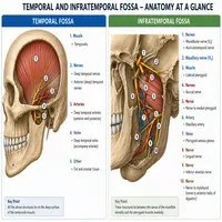
make a clear image for contents of temporal and infratemporal fossa
The skull hides several small but important spaces. Two of the most clinically useful are the temporal fossa and the infratemporal fossa. Together, the temporal and infratemporal fossa house muscles, nerves, and blood vessels that control chewing, jaw movement, and facial sensation. Students often confuse these two regions because they sit close together and share a border. However, each fossa has its own boundaries, contents, and clinical importance.
This guide breaks down the anatomy in plain language. You will find clear tables, a simple flowchart, and answers to common exam questions. By the end, you should understand exactly how these regions connect and why surgeons care about them so much.
What Is the Temporal Fossa?
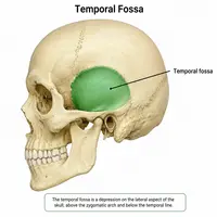
The temporal fossa sits on the side of the skull, just above the zygomatic arch. It forms a shallow depression that houses the temporalis muscle, one of the four major muscles of mastication. The fossa curves gently across the temporal bone, parietal bone, and part of the frontal bone.
Blood supply reaches this area mainly through branches of the maxillary artery. Nerve supply comes from the deep temporal nerves, which branch from the mandibular division of the trigeminal nerve. Because the temporal fossa lies just under the skin, it becomes an important landmark during scalp surgery and craniotomy planning.
What Is the Infratemporal Fossa?
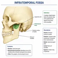
The infratemporal fossa lies below the temporal fossa, tucked behind the maxilla and deep to the mandible’s ramus. This space acts as a busy junction point. Several nerves, arteries, and muscles pass through it on their way to other regions of the head and neck.
Key structures inside the infratemporal fossa include the maxillary artery, pterygoid venous plexus, mandibular nerve branches, and the lateral and medial pterygoid muscles. Surgeons frequently approach this fossa during tumor removal, dental implant procedures, and management of jaw fractures.
Boundaries at a Glance
Clear boundaries help separate these two regions. The table below summarizes them side by side.
| Feature | Temporal Fossa | Infratemporal Fossa |
|---|---|---|
| Location | Above zygomatic arch | Below zygomatic arch |
| Roof | Superior temporal line | Infratemporal surface of greater wing of sphenoid |
| Floor | Not applicable (open above) | Medial pterygoid plate, lateral pterygoid |
| Anterior wall | Frontal process of zygomatic bone | Posterior surface of maxilla |
| Posterior wall | Not well defined | Tympanic plate, mastoid process |
| Medial wall | Not applicable | Lateral pterygoid plate |
| Lateral wall | Temporal fascia | Ramus of mandible |
Contents and Structures Passing Through
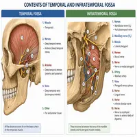
Both fossae carry structures that are vital for jaw movement and sensation. Here is a quick breakdown.
Temporal fossa contents:
- Temporalis muscle
- Deep temporal nerves and vessels
- Temporal fascia covering the muscle
Infratemporal fossa contents:
- Lateral and medial pterygoid muscles
- Maxillary artery and its branches
- Pterygoid venous plexus
- Mandibular nerve and its divisions
- Chorda tympani nerve
- Otic ganglion
As a result, damage to this region can affect chewing, taste sensation, and even middle ear function. Therefore, dentists and ENT surgeons study this space closely before any procedure near the jaw joint.
How the Two Fossae Connect
A common exam question asks how the temporal and infratemporal fossa communicate. The two spaces link through a gap beneath the zygomatic arch, allowing structures like the temporalis tendon and neurovascular bundles to travel between them.
Here is a simple flow of that connection:
Temporal Fossa
      |
      v
Deep to Zygomatic Arch (Communication Point)
      |
      v
Infratemporal Fossa
      |
      v
Pterygomaxillary Fissure
      |
      v
Pterygopalatine Fossa
This chain shows why infection or trauma in one region can quickly spread to nearby areas. For instance, a dental abscess near the maxillary molars can travel through this pathway toward deeper structures.
Clinical Relevance
Doctors and dental surgeons pay close attention to the temporal and infratemporal fossa during several procedures. Below are a few practical examples.
- Local anesthesia: Dentists inject anesthetic near the infratemporal fossa to block the mandibular nerve before molar extractions.
- Temporomandibular joint (TMJ) disorders: Pain often radiates from the temporal fossa due to overuse of the temporalis muscle.
- Tumor spread: Nasopharyngeal cancers can extend into the infratemporal fossa through the pterygomaxillary fissure.
- Fracture management: Zygomatic arch fractures may compress structures within the temporal fossa, limiting jaw movement.
Because these regions carry major nerves and vessels, even small surgical errors can cause lasting complications. Consequently, detailed anatomical knowledge remains essential for safe practice.
Quick Comparison Table
| Aspect | Temporal Fossa | Infratemporal Fossa |
|---|---|---|
| Main muscle | Temporalis | Pterygoids |
| Main artery | Deep temporal branches | Maxillary artery |
| Main nerve | Deep temporal nerves | Mandibular nerve |
| Depth | Superficial | Deep |
| Clinical focus | Scalp surgery, TMJ pain | Dental anesthesia, tumor spread |
Conclusion
The temporal and infratemporal fossa form a connected system that supports jaw function, facial sensation, and blood supply to deep facial structures. The temporal fossa sits superficially and houses the temporalis muscle, while the infratemporal fossa lies deeper and carries major nerves and vessels toward the face and skull base. Understanding their boundaries and connections helps students, dentists, and surgeons plan safer procedures. With the tables and flowchart above, this once-confusing topic becomes much easier to visualize and remember.
Frequently Asked Questions
The zygomatic arch acts as the main dividing landmark between the two spaces.
The mandibular division of the trigeminal nerve supplies most structures in this region.
Dentists use this space as a target point for nerve blocks during lower jaw procedures.
Yes, infection can travel through the communication point beneath the zygomatic arch.
The temporalis muscle fills most of the temporal fossa and helps close the jaw.