The neck holds many vital structures in a small space. To study this region clearly, anatomists divide the neck into smaller regions. These regions are called triangles. Each triangle has its own borders, contents, muscles, and nerves.
Dental students must understand this anatomy well. Procedures like nerve blocks, surgical extractions, and head and neck examinations depend on this knowledge. This guide breaks down each triangle in a simple, organized way. You will find tables, a flowchart, and clear explanations for every structure.
What Are Neck Triangles?
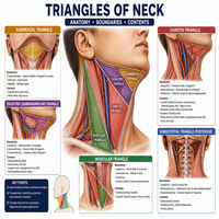
The neck is divided by the sternocleidomastoid muscle into two main regions: the anterior triangle and the posterior triangle. Each main triangle further splits into smaller triangles. This classification helps surgeons locate structures quickly during procedures.
Below is a simple flowchart that shows how the neck triangles are organized.
                         NECK
                          |
        -----------------------------------
        |                                 |
  Anterior Triangle                 Posterior Triangle
        |                                 |
 -------------------               -------------------
 |     |      |     |              |                 |
Submental  Digastric  Muscular  Carotid     Occipital  Subclavian
Triangle   Triangle   Triangle  Triangle    Triangle   Triangle
The suboccipital triangle sits separately, deep in the upper back of the neck. It belongs to a different region altogether. We will cover it later in this guide.
This basic classification of neck triangles makes the entire region easier to remember. Once you know the borders, the contents become easy to predict.
Anterior Triangle of Neck
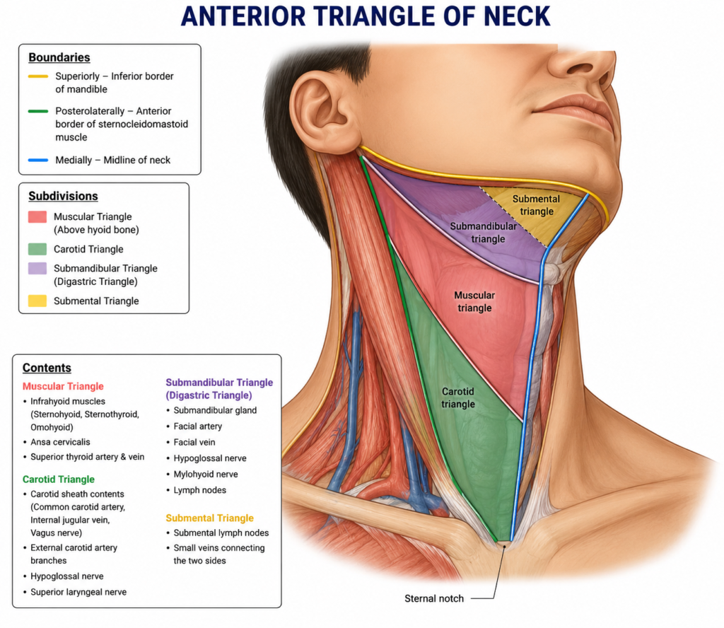
The anterior triangle of neck lies on the front side of the neck. It is bounded by clear landmarks, which make it simple to identify during dissection or examination.
| Feature | Description |
|---|---|
| Anterior border | Midline of the neck |
| Posterior border | Anterior edge of sternocleidomastoid |
| Superior border | Lower border of the mandible |
| Apex | Sternal notch |
| Roof | Skin, superficial fascia, platysma |
| Floor | Pharynx, larynx, thyroid gland |
This large triangle further divides into four smaller triangles: the submental, digastric, muscular, and carotid triangles. Each smaller triangle has unique contents. Therefore, learning them one by one makes the topic far less confusing.
Submental Triangle
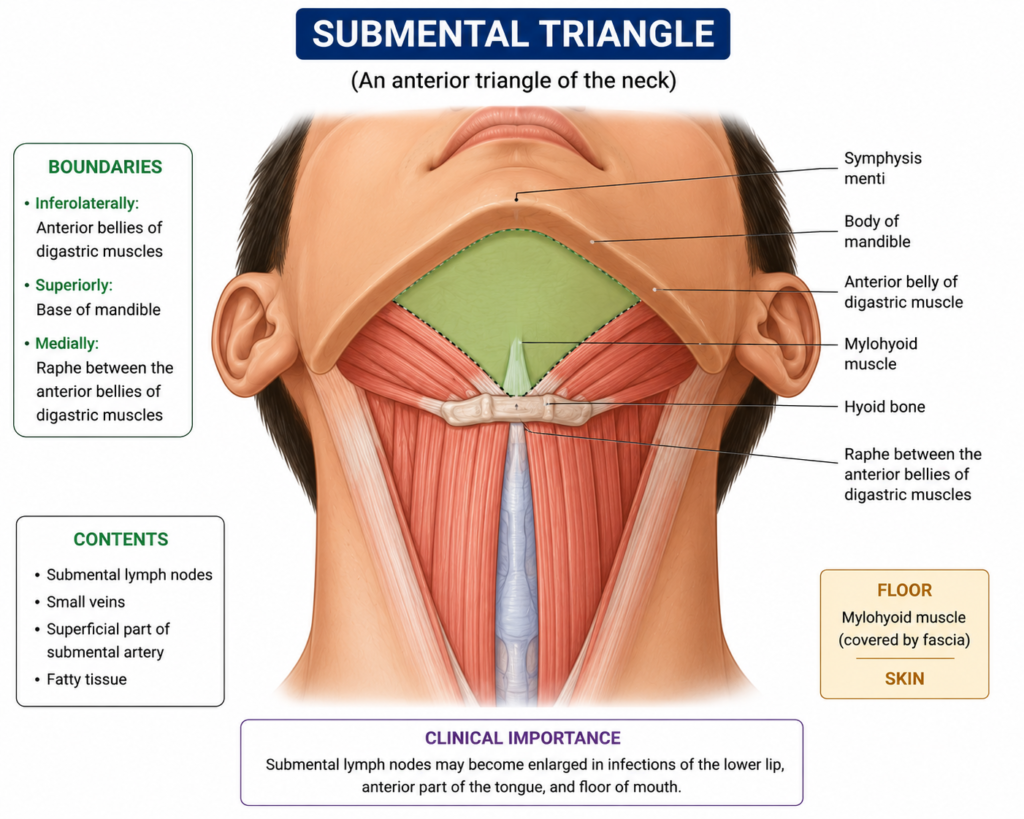
The submental triangle sits just below the chin. It is an unpaired triangle, meaning it does not have a left and right side. Instead, it forms a single space in the midline.
| Feature | Description |
|---|---|
| Boundaries | Hyoid bone (base), anterior bellies of digastric muscle (sides) |
| Floor | Mylohyoid muscle |
| Contents | Submental lymph nodes, small veins that form the anterior jugular vein |
| Nerve supply | Mylohyoid nerve (branch of inferior alveolar nerve) supplies the floor |
Dentists often check this area during oral cancer screenings. Submental lymph nodes can swell due to infections in the lower lip, chin, or floor of the mouth. As a result, this triangle holds real clinical value for dental practice.
Digastric Triangle
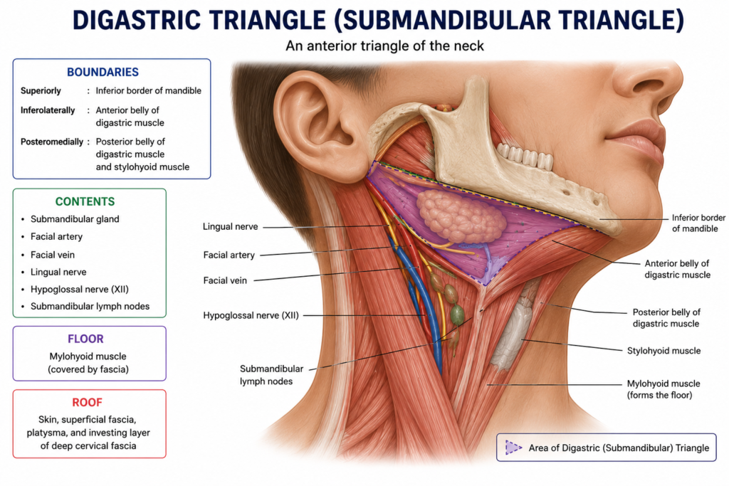
The digastric triangle, also called the submandibular triangle, lies just below the jaw. Many dental procedures, including submandibular gland assessment, relate directly to this region.
| Feature | Description |
|---|---|
| Boundaries | Body of mandible (base), anterior belly of digastric (front), posterior belly of digastric and stylohyoid (back) |
| Floor | Mylohyoid and hyoglossus muscles |
| Contents | Submandibular gland, submandibular lymph nodes, facial artery and vein, hypoglossal nerve, mylohyoid nerve and vessels |
| Nerve supply | Mandibular and cervical branches of the facial nerve supply nearby muscles |
The submandibular gland fills most of this space. Consequently, swelling in this triangle often points to gland infection or stone formation. Dental clinicians frequently palpate this area before oral surgery.
Muscular Triangle and Infrahyoid Muscles
The muscular triangle lies below the hyoid bone, closer to the midline. This triangle contains the infrahyoid muscles, which control the position of the larynx and hyoid bone.
| Feature | Description |
|---|---|
| Boundaries | Midline of neck (front), superior belly of omohyoid (above), anterior border of sternocleidomastoid (side) |
| Contents | Thyroid gland, larynx, trachea, infrahyoid muscles |
| Infrahyoid muscles | Sternohyoid, sternothyroid, thyrohyoid, omohyoid |
| Nerve supply | Ansa cervicalis (a loop formed by C1–C3 nerve fibers) |
These infrahyoid muscles depress the hyoid bone and larynx during swallowing and speech. Meanwhile, the thyroid gland sits centrally, making this triangle important during thyroid examinations and surgeries.
Carotid Triangle
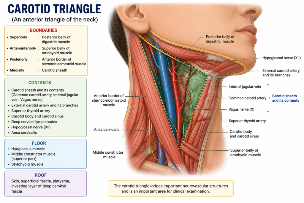
The carotid triangle is perhaps the most clinically significant of all neck triangles. It houses major blood vessels that supply the head and neck.
| Feature | Description |
|---|---|
| Boundaries | Posterior belly of digastric (above), superior belly of omohyoid (below), anterior border of sternocleidomastoid (back) |
| Contents | Common carotid artery, internal carotid artery, external carotid artery, internal jugular vein, vagus nerve, hypoglossal nerve |
| Nerve supply | Branches of the cervical plexus and vagus nerve |
The common carotid artery divides within this triangle into the internal and external carotid artery. The internal carotid artery supplies the brain, while the external carotid artery supplies the face and neck structures. Dentists must understand this division well, since several branches of the external carotid artery, such as the facial and maxillary arteries, directly supply oral and dental tissues.
Additionally, the vagus nerve and hypoglossal nerve pass through this region. Any injury here can affect swallowing, speech, or tongue movement. For this reason, surgeons handle this triangle with extra caution.
Suboccipital Triangle and Muscles
The suboccipital triangle lies deep in the back of the neck, below the occipital bone. Unlike the previous triangles, it belongs to the posterior neck region and is formed entirely by muscles.
| Feature | Description |
|---|---|
| Boundaries | Rectus capitis posterior major (above-medial), obliquus capitis superior (above-lateral), obliquus capitis inferior (below-lateral) |
| Floor | Posterior atlanto-occipital membrane, posterior arch of atlas |
| Contents | Vertebral artery, suboccipital nerve (dorsal ramus of C1) |
| Muscles forming the triangle | Rectus capitis posterior major, obliquus capitis superior, obliquus capitis inferior |
The suboccipital nerve supplies these muscles and helps control head movement. The vertebral artery, on the other hand, travels through this space before entering the skull. Therefore, this small triangle plays a big role in head posture and blood supply to the brain.
Quick Comparison Table of All Neck Triangles
| Triangle | Location | Key Contents |
|---|---|---|
| Submental | Below chin | Submental lymph nodes |
| Digastric | Below jaw | Submandibular gland |
| Muscular | Lower midline | Thyroid, infrahyoid muscles |
| Carotid | Upper lateral neck | Carotid arteries, vagus nerve |
| Suboccipital | Back of upper neck | Vertebral artery, suboccipital nerve |
This table offers a fast revision tool. Beginners can use it to recall locations and contents before exams or clinical rounds.
Conclusion
Understanding neck triangles helps dental students grasp the bigger picture of head and neck anatomy. Each triangle has its own boundaries, muscles, nerves, and vessels. Together, they form a complete map of this complex region.
Once you learn the borders of one triangle, the rest follow a similar pattern. Practice with diagrams, repeat the tables above, and review real specimens whenever possible. Over time, this anatomy becomes second nature, supporting safer and more confident dental practice.
Frequently Asked Questions
Neck triangles help dentists locate lymph nodes, glands, and blood vessels during examinations. They also guide safe injection sites and surgical planning.
The carotid triangle contains major blood vessels like the common carotid artery and external carotid artery. The muscular triangle, however, contains the thyroid gland and infrahyoid muscles.
The suboccipital nerve, which is the dorsal ramus of the first cervical nerve, supplies the muscles of this triangle.
This triangle houses the submandibular gland and related lymph nodes. Dentists often examine this area for swelling, infection, or stones before oral procedures.
The anterior triangle divides into four smaller triangles: the submental, digastric, muscular, and carotid triangles.