Bacterial Morphology, Identification, Genetics, and Taxonomy Explained
Bacteria are tiny, but their shapes tell a big story. A lab technician can often guess a genus just by looking through a microscope. That skill starts with one core idea: the morphology of bacteria.
This article breaks down bacterial shapes, arrangements, identification methods, genetic tools, and classification systems. Everything here is written for students, lab professionals, and curious readers who want a clear, updated guide to pathology basics.
What Is Bacterial Morphology?
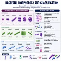
Morphology refers to the shape, size, and structural arrangement of a cell. In pathology, this single feature guides the first step of diagnosis. Doctors and lab techs rely on it before any advanced test begins.
Three basic shapes dominate the bacterial world:
- Cocci – round or spherical cells
- Bacilli – rod-shaped cells
- Spirilla – spiral or curved cells
Some organisms don’t fit neatly into these categories. Pleomorphic bacteria change shape depending on growth conditions. Mycoplasma is a classic example, since it lacks a rigid cell wall.
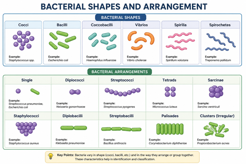
Common Bacterial Shapes and Arrangements
| Shape | Arrangement | Example Organism |
|---|---|---|
| Cocci | Single | Micrococcus |
| Cocci | Pairs (diplococci) | Neisseria gonorrhoeae |
| Cocci | Chains (streptococci) | Streptococcus pyogenes |
| Cocci | Clusters (staphylococci) | Staphylococcus aureus |
| Bacilli | Single rods | Escherichia coli |
| Bacilli | Chains | Bacillus cereus |
| Spirilla | Rigid spirals | Spirillum minus |
| Spirochetes | Flexible spirals | Treponema pallidum |
Arrangement matters as much as shape. Cells that stay in pairs, chains, or clusters after division often hint at specific genera. A trained eye combines shape and arrangement to narrow down possibilities fast.
Cell Structure Behind the Shape
The rigid cell wall, mainly made of peptidoglycan, holds a bacterium’s shape steady. Gram-positive species have a thick peptidoglycan layer. Gram-negative species carry a thinner layer wrapped in an outer membrane.
This structural difference forms the basis of the Gram stain, still one of the fastest identification tools in any lab. Within minutes, a technician learns whether an organism is gram-positive or gram-negative, plus its basic shape.
Flagella, capsules, and spores also shape how a cell behaves and survives. Spores, for instance, let genera like Bacillus and Clostridium withstand heat and drying. These features add extra clues during identification.
Methods Used for Identification of Bacteria
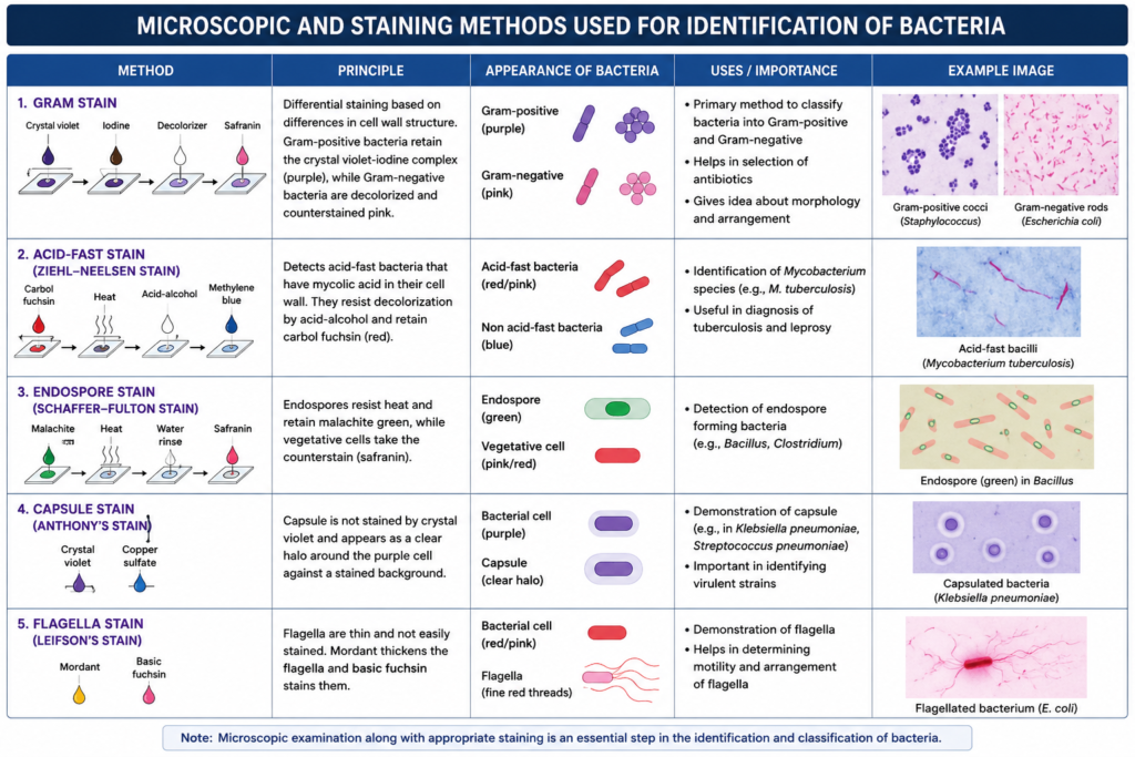
Shape alone rarely confirms a species. Labs combine several techniques to build a complete profile.
Microscopic and Staining Techniques
Gram staining remains the starting point in most clinical labs. Other stains serve specific purposes:
- Acid-fast stain – detects Mycobacterium species
- Endospore stain – reveals spore-forming genera
- Capsule stain – highlights encapsulated organisms like Klebsiella
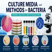
Culture-Based Methods
Growing bacteria on selective and differential media narrows down identity further. MacConkey agar separates lactose fermenters from non-fermenters. Blood agar reveals hemolytic patterns unique to certain streptococci.
Colony color, texture, smell, and growth pattern all add pieces to the puzzle. Experienced microbiologists often recognize a species just from colony appearance.
Biochemical Testing
Enzyme-based tests confirm metabolic traits. Catalase, oxidase, coagulase, and urease tests are routine in most diagnostic labs. Each test targets a specific biochemical pathway, and results get compared against known reference charts.
Molecular and Genetic Identification
Modern labs increasingly rely on genetic tools instead of, or alongside, traditional methods. Polymerase chain reaction (PCR) amplifies specific DNA sequences for rapid confirmation. 16S rRNA gene sequencing has become a gold standard for identifying bacteria that resist normal culturing.
These tools cut diagnosis time from days to hours. They also catch organisms that grow slowly or not at all on standard media.
Bacterial Identification Flowchart
Specimen Collected
        │
        ▼
   Gram Stain Performed
        │
   ┌────┴────┐
   ▼         ▼
Gram-Positive   Gram-Negative
   │                │
   ▼                ▼
Culture on Media   Culture on Media
   │                │
   ▼                ▼
Biochemical Tests   Biochemical Tests
   │                │
   └────┬───────────┘
        ▼
  Molecular Confirmation
     (PCR / Sequencing)
        │
        ▼
   Final Species Identification
This stepwise process keeps labs consistent and accurate. Each stage filters out possibilities until one identity remains.
Genetics of Bacteria: The Blueprint Behind Behavior
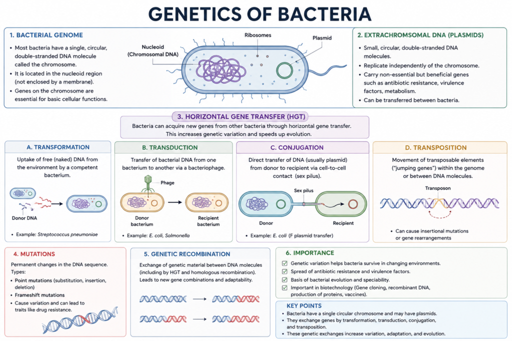
Bacterial genetics explains why some strains cause disease while others stay harmless. A single circular chromosome carries most genetic material, but plasmids often carry extra genes for antibiotic resistance or toxin production.
Horizontal gene transfer spreads traits between bacteria without reproduction. Three main mechanisms drive this process:
- Transformation – uptake of free DNA from the environment
- Transduction – gene transfer through bacteriophages
- Conjugation – direct DNA transfer through cell-to-cell contact
These mechanisms explain how antibiotic resistance jumps between unrelated species so quickly. A resistant gene in one organism can end up in a completely different genus within days.
Mutations also drive evolution at the genetic level. Even small changes in a single gene can alter virulence, resistance, or metabolic ability. Genetic study connects directly back to morphology, since certain genes control cell wall synthesis and shape.
Taxonomy and Classification of Bacteria
Taxonomy organizes the bacterial world into a logical hierarchy. Classification of bacteria groups organisms by shared traits, going from broad domains down to specific species.
The Taxonomic Hierarchy
| Rank | Example |
|---|---|
| Domain | Bacteria |
| Phylum | Proteobacteria |
| Class | Gammaproteobacteria |
| Order | Enterobacterales |
| Family | Enterobacteriaceae |
| Genus | Escherichia |
| Species | Escherichia coli |
Modern taxonomy no longer relies only on shape or staining. Genetic sequencing reshaped classification of bacteria over the past two decades. The 16S rRNA gene, present in nearly all bacteria, now serves as a universal marker for grouping related organisms.
Systems Used for Classification of Bacteria
Several systems guide how scientists name and group organisms:
- Numerical taxonomy – compares multiple traits using statistical scoring
- Phylogenetic taxonomy – groups organisms by evolutionary relationships
- Polyphasic taxonomy – combines genetic, chemical, and phenotypic data
Polyphasic taxonomy currently dominates formal species description. It blends traditional morphology of bacteria with molecular data, giving a fuller picture than either method alone.
Why This Matters in Pathology
Correct identification changes treatment decisions directly. A wrongly identified organism can lead to ineffective antibiotics or delayed care. Fast, accurate classification of bacteria also supports outbreak tracking and public health responses.
Hospitals now use rapid diagnostic panels that combine morphology, biochemical data, and genetic markers in a single test. This shift toward integrated diagnostics reflects how far pathology has advanced beyond the microscope alone.
Conclusion
The morphology of bacteria still forms the foundation of clinical microbiology, even as genetic tools take a larger role. Shape, staining, and arrangement guide the first clues. Biochemical tests and molecular sequencing confirm the final answer.
Genetics explains how bacteria adapt, resist treatment, and evolve over time. Taxonomy ties everything together, giving every organism a clear place within a shared classification system. Together, these fields give pathologists a complete, reliable path from sample to diagnosis.
Understanding this full picture helps students, lab professionals, and healthcare workers make faster, more confident decisions in real clinical settings.
Frequently Asked Questions
Morphology refers to the shape, size, and cellular arrangement of a bacterium, such as cocci, bacilli, or spirilla. It is often the first clue used during identification
Gram staining separates bacteria into gram-positive and gram-negative groups based on cell wall structure. This quick test guides which further tests a lab should run next.
Genetic sequencing, especially of the 16S rRNA gene, reveals evolutionary relationships that shape and staining alone cannot show. This has made classification of bacteria far more accurate than older methods.
It is a process where bacteria share genetic material without reproducing, through transformation, transduction, or conjugation. This process explains how antibiotic resistance spreads rapidly between species.
Polyphasic taxonomy combines morphological, biochemical, and genetic data to classify bacteria. It is currently the standard approach for formally describing new bacterial species.