Osteology of the Head and Neck
Osteology of the Head and Neck is a branch of anatomy that deals with the study of the bones of the skull, face, and neck. It provides a detailed understanding of the shape, structure, and function of these bones. Knowledge of Osteology of the Head and Neck is essential for medical, dental, and surgical students because it forms the foundation for understanding craniofacial anatomy, trauma, and surgical procedures. Moreover, it helps clinicians identify anatomical landmarks and diagnose fractures or congenital abnormalities. Therefore, mastering Osteology of the Head and Neck is important for both academic learning and clinical practice.
Bones of the Skull
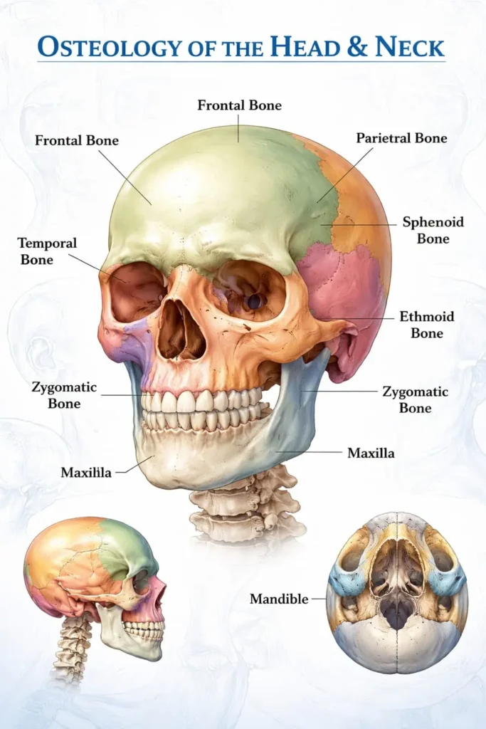
The adult skull consists of 22 bones, which are divided into the cranial bones (8) and the facial bones (14). The cranial bones protect the brain, whereas the facial bones provide the framework of the face and support the eyes, nose, and mouth. Furthermore, these bones are connected by sutures that provide strength while allowing slight flexibility during development. Osteology of the Head and Neck explains how these bones work together to protect vital organs and support facial structures.
Skull Joints
Most skull bones are connected by fibrous joints called sutures. These joints are immovable in adults and provide stability to the skull. The major sutures include the coronal, sagittal, lambdoid, and squamous sutures. However, the temporomandibular joint (TMJ) is a synovial joint that permits movement of the mandible during speaking and chewing. This joint is one of the most clinically significant structures in Osteology of the Head and Neck.
Anatomical Position
The skull is studied in the standard anatomical position, where the head is upright and the Frankfurt horizontal plane is parallel to the ground. In this position, the inferior margin of the orbit and the upper border of the external auditory meatus lie in the same horizontal plane. Consequently, this position allows accurate identification of anatomical landmarks.
Exterior of the Skull
The exterior of the skull is divided into different views known as normae. These views include the superior, posterior, anterior, lateral, and inferior aspects. Each view demonstrates unique anatomical features that are important for identifying bones, sutures, foramina, and muscle attachments. Therefore, studying the external skull is a key component of Osteology of the Head and Neck.
Norma Verticalis
The Norma Verticalis represents the superior view of the skull. It mainly shows the frontal bone, parietal bones, and occipital bone. The coronal, sagittal, and lambdoid sutures are clearly visible from this aspect. Additionally, the bregma and lambda serve as important anatomical landmarks for clinicians and surgeons.
Norma Occipitalis
The Norma Occipitalis is the posterior view of the skull. It mainly consists of the occipital bone along with parts of the parietal and temporal bones. The external occipital protuberance and superior nuchal lines provide attachment sites for neck muscles. As a result, this region plays an important role in supporting head movements.
Norma Frontalis
The Norma Frontalis is the anterior view of the skull. It displays the frontal bone, nasal bones, maxillae, zygomatic bones, mandible, and orbital cavities. Moreover, the piriform aperture forms the entrance to the nasal cavity. This view is essential for evaluating facial symmetry and identifying facial fractures.
Attachments and Structures Passing Through Foramina
Numerous muscles, ligaments, nerves, and blood vessels are associated with the skull. Foramina are openings that allow important structures to pass between the cranial cavity and surrounding regions. For example, the optic canal transmits the optic nerve, while the foramen rotundum transmits the maxillary nerve. Therefore, understanding these openings is vital in Osteology of the Head and Neck.
Norma Lateralis
The Norma Lateralis presents the side view of the skull. It includes the temporal fossa, zygomatic arch, external acoustic meatus, mastoid process, and styloid process. Furthermore, the pterion, where four bones meet, is an important clinical landmark because it overlies the middle meningeal artery.
Emissary Veins
Emissary veins connect the veins outside the skull with the intracranial venous sinuses. Although they help equalize venous pressure, they may also provide a pathway for infections to spread into the cranial cavity. Consequently, these veins have significant clinical importance.
Norma Basalis
The Norma Basalis is the inferior view of the skull. It is divided into anterior, middle, and posterior parts. This aspect contains numerous foramina through which cranial nerves and blood vessels enter or leave the skull. In addition, several muscles of the neck and pharynx attach to this region.
Anterior Part
The anterior part includes the hard palate, alveolar arches, and posterior nasal apertures. It forms the roof of the oral cavity and the floor of the nasal cavity. Therefore, it is essential for speech, swallowing, and mastication.
Middle Part
The middle part contains the sphenoid bone, temporal bones, carotid canal, and foramen ovale. These structures allow the passage of major nerves and arteries supplying the brain and face.
Posterior Part
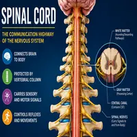
The posterior part is dominated by the occipital bone and the foramen magnum. The spinal cord passes through the foramen magnum to connect with the brain. Additionally, this region contains the occipital condyles, which articulate with the atlas vertebra.
Interior of the Skull
The interior of the skull houses the brain and its protective coverings. It contains grooves for arteries, venous sinuses, and cranial nerves. Moreover, the cranial cavity is divided into three cranial fossae that support different parts of the brain.
Diploic Veins
Diploic veins are located within the diploë, the spongy bone between the inner and outer tables of the skull. They drain blood from the skull bones and communicate with emissary veins. Consequently, infections may spread through these venous channels.
Cranial Vault
The cranial vault forms the upper part of the skull and protects the cerebral hemispheres. It consists mainly of the frontal, parietal, and occipital bones. Furthermore, its curved shape helps distribute the force of external impacts.
Base of the Skull
The base of the skull supports the brain and contains numerous foramina for cranial nerves and blood vessels. It is structurally complex because several bones contribute to its formation. Therefore, fractures of the skull base may damage vital neurovascular structures.
Anterior Cranial Fossa
The anterior cranial fossa supports the frontal lobes of the brain. It is formed by the frontal, ethmoid, and sphenoid bones. The cribriform plate allows olfactory nerve fibers to pass into the nasal cavity.
Middle Cranial Fossa
The middle cranial fossa accommodates the temporal lobes and the pituitary gland. It contains important foramina, including the superior orbital fissure, foramen rotundum, foramen ovale, and foramen spinosum. These openings transmit several cranial nerves and arteries.
Posterior Cranial Fossa
The posterior cranial fossa is the deepest cranial compartment. It contains the cerebellum, pons, and medulla oblongata. Furthermore, the internal acoustic meatus and jugular foramen transmit essential nerves and blood vessels.
Connections of Parasympathetic Ganglia
Parasympathetic ganglia in the head include the ciliary, pterygopalatine, submandibular, and otic ganglia. These ganglia receive fibers from cranial nerves and supply glands of the head. Consequently, they regulate tear production, salivation, and other secretory functions.
Attachments on the Interior of the Skull
The dura mater is firmly attached to specific areas inside the skull. In addition, structures such as the falx cerebri and tentorium cerebelli are anchored to the cranial bones, providing support to the brain.
The Orbit
The orbit is a pyramidal cavity that houses the eyeball and its associated structures. It is formed by seven bones: frontal, sphenoid, zygomatic, maxilla, lacrimal, ethmoid, and palatine. Moreover, it protects the eye while allowing its movement.
Foramina in Relation to the Orbit
Several foramina communicate with the orbit. The optic canal transmits the optic nerve and ophthalmic artery, whereas the superior orbital fissure allows the passage of cranial nerves controlling eye movements. Additionally, the inferior orbital fissure transmits vessels and nerves to the face.
Foetal Skull
The foetal skull differs from the adult skull because it contains fontanelles and unfused sutures. These flexible areas facilitate childbirth and permit rapid brain growth during infancy. Eventually, the fontanelles close, and the sutures become firmly united.
Conclusion
In conclusion, Osteology of the Head and Neck provides a comprehensive understanding of the bones, joints, foramina, cranial fossae, and orbital structures of the skull. Moreover, this knowledge is fundamental for dentistry, medicine, neurosurgery, and maxillofacial surgery. By understanding the anatomical relationships of the skull, healthcare professionals can diagnose diseases, manage trauma, and perform surgical procedures more effectively. Therefore, Osteology of the Head and Neck remains one of the most important topics in human anatomy and continues to play a vital role in clinical practice.