Cell Junctions: Definition, Types & Functions Explained
Healthy tissues, including the gums and oral mucosa, depend on cells that stick together, communicate, and form protective barriers. This teamwork happens through specialized structures known as cell junctions. Without these structures, tissues would simply fall apart, fluids would leak uncontrollably, and cells would lose the ability to “talk” to their neighbors. In this article, we explore the definition, classification, and clinical relevance of these essential structures, with a special focus on how dental and oral tissues rely on them every single day.
What Are Cell Junctions?
Cell junctions are specialized points of contact between adjacent cells, or between a cell and the surrounding extracellular matrix. These contact points hold tissues together, regulate the movement of substances between cells, and allow cells to coordinate their activities as a single functional unit. Because epithelial tissues, such as the lining of the oral cavity, face constant mechanical stress and exposure to bacteria, they depend heavily on strong, well-organized junctions.
Generally, cell junctions perform three core jobs: mechanical attachment, barrier formation, and intercellular communication. Therefore, scientists usually classify them according to which of these three jobs they perform best. Additionally, some junctions multitask, contributing to more than one function at once. As a result, understanding their classification makes it much easier to understand tissue physiology as a whole.
Classification of Cell Junctions
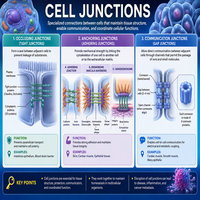
Cell junctions are broadly classified into three major categories based on function. The table below summarizes this classification clearly.
| Category | Primary Function | Common Examples |
|---|---|---|
| Occluding Junctions | Seal the space between cells, prevent leakage | Tight junctions (zonula occludens) |
| Anchoring Junctions | Provide mechanical strength, attach cells to each other or matrix | Desmosomes, adherens junctions, hemidesmosomes |
| Communication Junctions | Allow direct exchange of ions and small molecules | Gap junctions |
Although each category has a distinct role, they often work together within the same tissue. For instance, oral epithelial cells contain occluding junctions near their apical surface, anchoring junctions throughout the lateral membrane, and communication junctions scattered along the cell body. Consequently, a single epithelial cell may rely on all three junction types simultaneously to remain structurally sound and functionally active.
Simple Classification Flowchart
                     CELL JUNCTIONS
                          |
   -----------------------------------------------
   |                      |                      |
OCCLUDING            ANCHORING              COMMUNICATION
JUNCTIONS            JUNCTIONS                JUNCTIONS
   |                      |                      |
Tight Junctions     Desmosomes               Gap Junctions
                     Adherens Junctions
                     Hemidesmosomes
Occluding Junctions
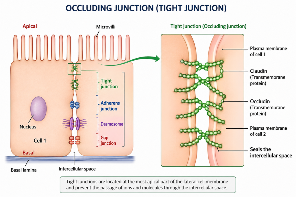
Occluding junctions, also called tight junctions, form a continuous seal between neighboring epithelial cells. Because this seal blocks the free passage of fluids and pathogens, it protects deeper tissue layers from harmful invasion. Tight junctions are typically located near the apical surface of epithelial cells, where they form a beltlike structure that encircles the entire cell.
Structurally, transmembrane proteins such as claudins and occludins interlock with similar proteins on the adjacent cell, sealing the intercellular space almost completely. Moreover, this sealing effect creates a polarity within the cell, separating the apical domain from the basolateral domain. Without this separation, selective absorption and secretion processes would become impossible.
In the oral cavity, occluding junctions in the gingival epithelium prevent bacteria and toxins from easily penetrating the underlying connective tissue. However, when these junctions become compromised, due to inflammation or chronic infection, bacterial products can infiltrate deeper tissues more easily, contributing to periodontal disease progression. Thus, the integrity of occluding junctions plays a direct role in maintaining periodontal health.
Anchoring Junctions
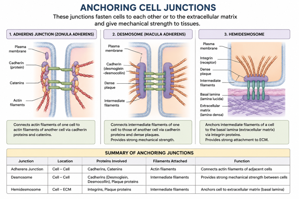
Anchoring junctions provide the mechanical glue that keeps tissues intact under stress. Unlike occluding junctions, their main purpose is not sealing but rather binding cells firmly to one another or to the underlying basement membrane. Three major types fall under this category: desmosomes, adherens junctions, and hemidesmosomes.
Desmosomes act like spot welds, linking the intermediate filaments of two neighboring cells through cadherin proteins. Similarly, adherens junctions connect actin filaments across the cell membrane, forming a continuous adhesive belt just below the tight junctions. Meanwhile, hemidesmosomes anchor epithelial cells to the basement membrane rather than to other cells, providing stability against shearing forces.
Because oral mucosa and gingival tissue endure constant chewing forces, friction from food, and exposure to temperature changes, anchoring junctions are particularly important here. For example, desmosomes in the gingival epithelium resist the mechanical stress generated during mastication. In addition, hemidesmosomes secure the epithelium to the connective tissue below, preventing detachment that could otherwise lead to ulceration or erosion. Consequently, any genetic or autoimmune disorder affecting desmosomal proteins, such as pemphigus vulgaris, often produces painful oral blistering and tissue breakdown.
Communication Junctions
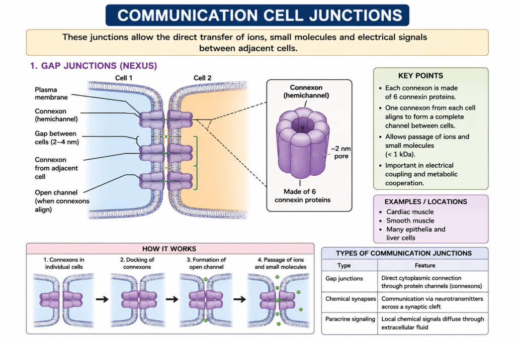
Communication junctions, commonly known as gap junctions, allow direct cytoplasmic continuity between adjacent cells. These structures consist of protein channels called connexons, which align precisely with connexons on the neighboring cell to form a functional channel. Through this channel, ions, small metabolites, and signaling molecules pass freely from one cell to another.
This direct communication enables coordinated cellular responses across an entire tissue. For instance, gap junctions allow rapid electrical signaling in cardiac muscle, ensuring synchronized contraction. Likewise, in epithelial tissues, gap junctions help coordinate responses to injury, inflammation, and growth signals. Furthermore, because the channels can open or close depending on intracellular calcium and pH levels, cells can regulate how much communication occurs at any given moment.
Within the periodontium, communication junctions support coordinated healing responses after minor trauma or surgical procedures. As a result, cells located far from a wound site can still receive signaling cues that promote a unified tissue repair process. Overall, gap junctions complete the functional picture established by occluding and anchoring junctions, since structural integrity alone is not enough without proper cellular communication.
Why Cell Junctions Matter in Dentistry
Dental professionals encounter the consequences of healthy or compromised cell junctions on a regular basis. Strong tight junctions reduce bacterial penetration through gingival epithelium, while robust desmosomes and hemidesmosomes protect against mucosal injury during everyday function. Meanwhile, functional gap junctions support faster, well-coordinated healing after extractions, periodontal therapy, or oral surgery.
Therefore, when clinicians assess oral tissue health, they are indirectly evaluating the performance of these microscopic structures. For example, signs of gingival inflammation often reflect disrupted occluding junctions, whereas blistering conditions point toward anchoring junction abnormalities. In this way, a basic understanding of physiology directly informs better clinical decision-making.
Conclusion
Cell junctions form the structural and functional backbone of every epithelial tissue, including those lining the oral cavity. Occluding junctions seal tissues and block harmful invaders, anchoring junctions provide mechanical strength against everyday wear, and communication junctions allow cells to coordinate their activities as a unified system. For dental professionals and students alike, understanding the physiology behind these structures provides valuable insight into both healthy function and the early signs of disease. Ultimately, a solid grasp of cell junctions supports better diagnosis, more informed treatment planning, and a deeper appreciation for how delicate, yet resilient, oral tissues truly are.
Frequently Asked Questions
Cell junctions hold cells together, create protective barriers, and allow neighboring cells to communicate directly with one another.
The three main types are occluding junctions, anchoring junctions, and communication junctions, each serving a distinct structural or functional role.
Tight junctions form a seal near the apical surface of epithelial cells, blocking bacteria and toxins from easily reaching deeper connective tissue layers.
Desmosomes provide mechanical attachment between cells using intermediate filaments, while gap junctions create channels that allow ions and small molecules to pass directly between cells.
Strong, intact junctions reduce bacterial penetration, support tissue resilience against chewing forces, and promote coordinated healing, all of which protect against periodontal disease.