Human life begins with a single cell, yet it eventually grows into a body with billions of specialized cells. General embryology studies this remarkable journey, from the moment two cells unite to the formation of organs and tissues.
Understanding this process matters for many healthcare fields, including dentistry. Therefore, many dental and medical professionals study early development to understand how facial structures, teeth, and the nervous system form. This article breaks the topic into clear stages: germ cell formation, fertilization, prenatal development, induction, competence, differentiation, the formation of the three-layered embryo, neural tube formation, and the fate of germ layers.
We will also include a flowchart and a comparison table to simplify these concepts. By the end, you will have a clear, practical understanding of general embryology and why it remains essential knowledge in biology and healthcare education.
Germ Cell Formation and Fertilization
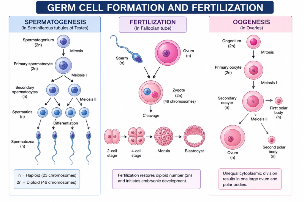
Germ cells, also called gametes, form through a special process called gametogenesis. In males, this process produces sperm cells. In females, it produces an egg cell, or ovum. Both cells carry half the genetic material needed to create a new individual.
During ovulation, the ovary releases a mature egg into the fallopian tube. Meanwhile, millions of sperm travel toward the egg after intercourse, though only one sperm typically succeeds in fertilizing it. Once a sperm penetrates the egg’s outer layer, fertilization occurs, and the two sets of chromosomes combine to form a single cell called a zygote.
This zygote contains the complete genetic blueprint for the new organism. Immediately afterward, the zygote begins dividing rapidly through a process called cleavage. As a result, the single cell becomes multiple smaller cells, eventually forming a solid ball called a morula.
The morula continues developing into a blastocyst, a structure containing an inner cell mass and a fluid-filled cavity. This inner cell mass eventually forms the embryo itself, while outer cells contribute to the placenta. Consequently, fertilization marks the true starting point of prenatal development.
Prenatal Development Overview
Prenatal development describes the entire process from fertilization to birth. It typically divides into three main periods: the germinal period, the embryonic period, and the fetal period.
The germinal period covers the first two weeks, including fertilization, cleavage, and blastocyst formation. Following this, the embryonic period spans weeks three through eight. During this stage, major organs and body systems begin forming rapidly.
Afterward, the fetal period continues from week nine until birth. During this time, organs mature, and the body grows significantly in size. Throughout all three periods, the body relies heavily on precise cell communication and timing.
Notably, the embryonic period is the most sensitive window for development. Even small disruptions during this stage can affect organ formation, including facial and dental structures. This is one reason general embryology remains highly relevant in dental education, since many craniofacial features form during these early weeks.
Induction and Competence
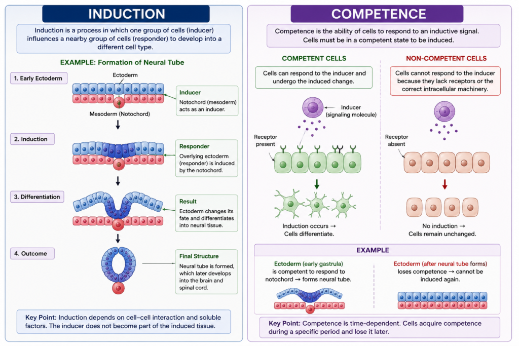
Induction is a process where one group of cells influences the development of a neighboring group. Essentially, signals from one tissue trigger specific changes in another tissue nearby.
For induction to succeed, the responding tissue must possess competence. Competence refers to a tissue’s ability to respond appropriately to an inductive signal. Without competence, even a strong signal will have no effect.
For example, the notochord sends signals that induce the overlying ectoderm to form the neural plate. However, this only works if the ectoderm is competent to receive that signal at the right developmental stage. Timing plays a critical role here, since competence often changes as development progresses.
This relationship between induction and competence ensures organized, predictable development. Similarly, many craniofacial and dental structures form through repeated rounds of induction between epithelial and mesenchymal tissues. Therefore, this concept connects directly to how teeth and jaw structures eventually take shape.
Differentiation
Differentiation is the process where unspecialized cells become specialized cells with specific structures and functions. Initially, embryonic cells appear similar to one another. Gradually, they begin expressing different genes, leading to distinct cell types.
This process depends heavily on gene expression patterns activated by signals from neighboring cells. Consequently, a single fertilized egg can eventually produce nerve cells, muscle cells, blood cells, and many other specialized types.
Differentiation does not happen randomly. Instead, it follows a tightly controlled sequence influenced by location, timing, and chemical signals. As a result, cells in one part of the embryo develop very differently from cells in another part, even though they originally shared identical genetic material.
This organized specialization ultimately builds every tissue and organ in the body, including the structures that form teeth, gums, and supporting bone.
Formation of the Three-Layered Embryo
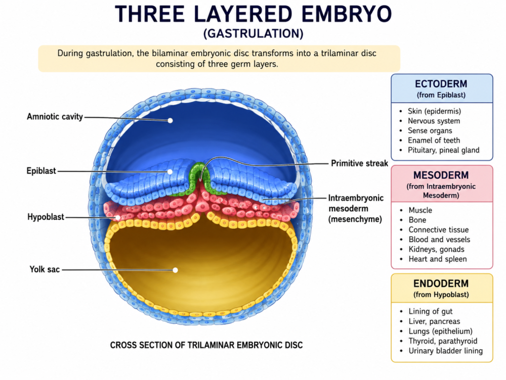
During the third week of development, the embryo undergoes gastrulation, a process that creates three distinct germ layers. These layers are the ectoderm, mesoderm, and endoderm.
This transformation begins when cells migrate inward through a structure called the primitive streak. Afterward, these migrating cells form the middle layer, known as the mesoderm. Meanwhile, the outer layer becomes the ectoderm, and the inner layer becomes the endoderm.
Flowchart: From Fertilization to Three Germ Layers
              Fertilization (Zygote Formed)
                          |
                          v
                Cleavage and Morula Stage
                          |
                          v
                  Blastocyst Formation
                          |
                          v
                 Gastrulation Begins
                          |
                          v
        Three Germ Layers Form: Ectoderm,
              Mesoderm, and Endoderm
Each layer eventually gives rise to specific tissues and organs throughout the body, which we will explore in the final section.
Formation of the Neural Tube
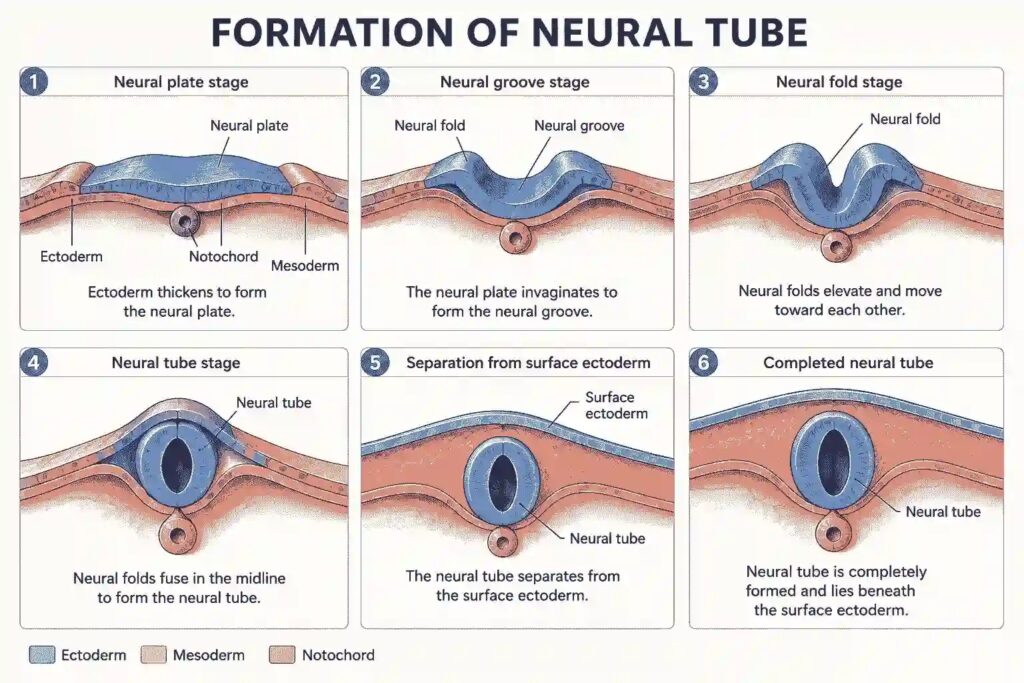
Shortly after gastrulation, the ectoderm thickens above the notochord to form the neural plate. Soon afterward, the edges of this plate rise upward, forming neural folds. These folds eventually meet and fuse along the midline, creating the neural tube.
This neural tube becomes the foundation of the entire central nervous system, including the brain and spinal cord. Notably, failure of proper fusion can lead to serious developmental conditions, such as neural tube defects.
Nutrition plays a major role during this stage. For instance, adequate folic acid intake significantly reduces the risk of neural tube defects. Therefore, prenatal care often emphasizes proper nutrition during these early, critical weeks of pregnancy.
Fate of Germ Layers
Each of the three germ layers eventually develops into specific structures throughout the body. Understanding this fate helps explain how complex systems originate from simple early layers.
| Germ Layer | Major Derivatives |
|---|---|
| Ectoderm | Skin, nervous system, tooth enamel, hair |
| Mesoderm | Muscles, bones, blood vessels, dentin |
| Endoderm | Digestive lining, lungs, liver, pancreas |
Interestingly, both ectoderm and mesoderm contribute directly to dental structures, since enamel forms from ectoderm while dentin and supporting bone arise from mesoderm. This connection highlights why general embryology holds particular relevance for dental professionals studying craniofacial development.
Conclusion
General embryology traces an extraordinary path from a single fertilized cell to a fully formed human body. Through fertilization, prenatal development, induction, competence, and differentiation, the embryo gradually organizes itself into three distinct germ layers. These layers then give rise to essential structures, including the neural tube and the tissues that form teeth and jaws.
This knowledge offers valuable insight for anyone studying biology, medicine, or dentistry. In short, every organ, tissue, and structure in the body traces its origin back to these early, carefully coordinated developmental events.
Frequently Asked Questions
General embryology is the study of how an organism develops from fertilization through prenatal stages until birth, including organ and tissue formation.
Induction is a signal sent by one tissue to influence another, while competence is the responding tissue’s ability to react appropriately to that signal.
Gastrulation creates the three germ layers, ectoderm, mesoderm, and endoderm, which later form all body tissues and organs.
The neural tube forms when the edges of the neural plate rise, fold inward, and fuse along the midline during early development.
Tooth enamel forms from ectoderm, while dentin and supporting bone arise from mesoderm, linking early germ layer formation directly to dental structures.