Embryology of Head, Face, and Neck: A Simple Guide
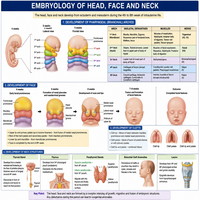
Human development begins with a few simple cell layers. From these layers, complex structures eventually form. The embryology of head, face, and neck explains how this transformation happens.
This process shapes the skull, jaws, muscles, and facial features. Dentists need this knowledge deeply. Why? Because most craniofacial anomalies originate during early embryonic stages.
Understanding the embryology of head, face, and neck also helps explain birth defects like cleft lip and palate. So, let’s break this topic into simple, clear steps.
This guide covers pharyngeal arches, facial development, neck formation, and clinical applications. Additionally, we will look at common developmental anomalies.
Pharyngeal Arches: The Foundation
Pharyngeal arches appear early in development. These paired structures form the basic framework of the head and neck.
Formation Timeline
Pharyngeal arches develop around the fourth week of gestation. Six arches form initially, though the fifth arch disappears quickly. Each arch contains specific components.
| Component | Description |
|---|---|
| Cartilage bar | Forms skeletal elements |
| Muscle component | Forms specific muscle groups |
| Nerve | Supplies the arch and its derivatives |
| Artery | Supplies blood to arch structures |
Major Arches and Their Derivatives
| Arch | Nerve | Major Derivatives |
|---|---|---|
| First (Mandibular) | Trigeminal (V) | Mandible, muscles of mastication |
| Second (Hyoid) | Facial (VII) | Stapes, styloid process, muscles of facial expression |
| Third | Glossopharyngeal (IX) | Greater horn of hyoid bone |
| Fourth and Sixth | Vagus (X) | Laryngeal cartilages, pharyngeal muscles |
The first arch matters most for dentistry. It forms the mandible and maxillary processes. Consequently, problems here often cause jaw and facial deformities.
Development of the Face
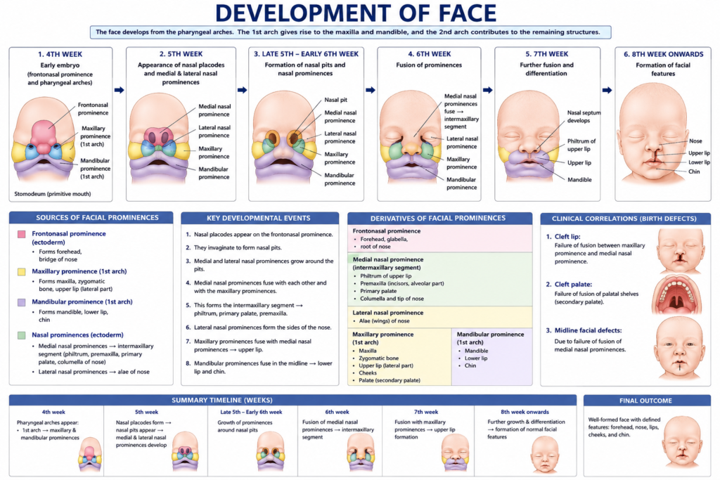
Facial development involves multiple swellings called processes. These processes grow and fuse together gradually.
Key Facial Processes
- Frontonasal process – Forms forehead and nose
- Maxillary processes – Form upper jaw and lateral lip
- Mandibular processes – Form lower jaw and lip
Here’s a simple flowchart of facial formation:
Frontonasal Process
        |
        ▼
Forms Nasal Placodes
        |
        ▼
Medial & Lateral Nasal Processes Form
        |
        ▼
Fusion with Maxillary Processes
        |
        ▼
Upper Lip and Nose Complete
Meanwhile, mandibular processes fuse in the midline early. This fusion forms the lower lip and chin. Therefore, mandibular fusion defects are rarer than upper lip defects.
Nasal placodes also play a crucial role. They form nasal pits, which later become nostrils. Additionally, these structures contribute to primary palate formation.
Development of the Neck
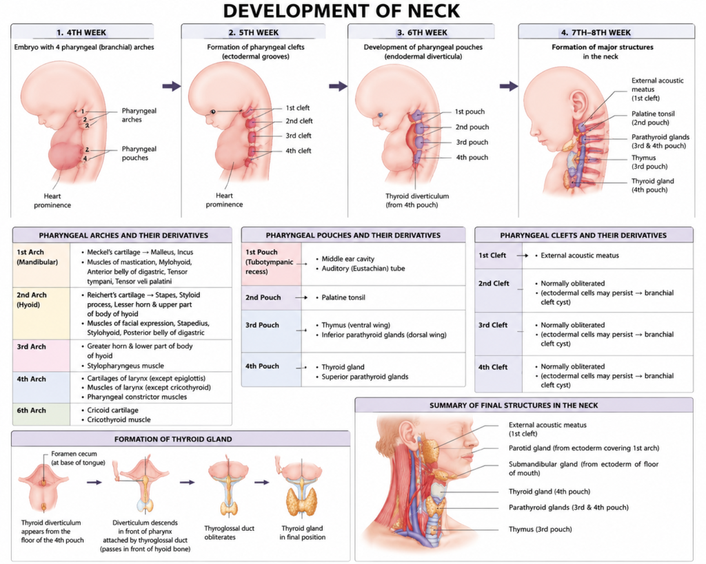
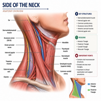
Neck development relates closely to pharyngeal arch formation. Pharyngeal pouches and clefts contribute significantly to this region.
Pharyngeal Pouches
These internal pouches form important glandular structures.
| Pouch | Derivative |
|---|---|
| First | Eustachian tube, middle ear cavity |
| Second | Palatine tonsil |
| Third | Inferior parathyroid, thymus |
| Fourth | Superior parathyroid |
Pharyngeal Clefts
External clefts mostly disappear during development. However, the first cleft persists and forms the external auditory meatus. Remaining clefts get buried by overgrowth of the second arch.
Notably, incomplete obliteration of clefts can cause branchial cysts or fistulas. These anomalies often appear along the neck’s lateral border in children.
Development of Tongue and Palate
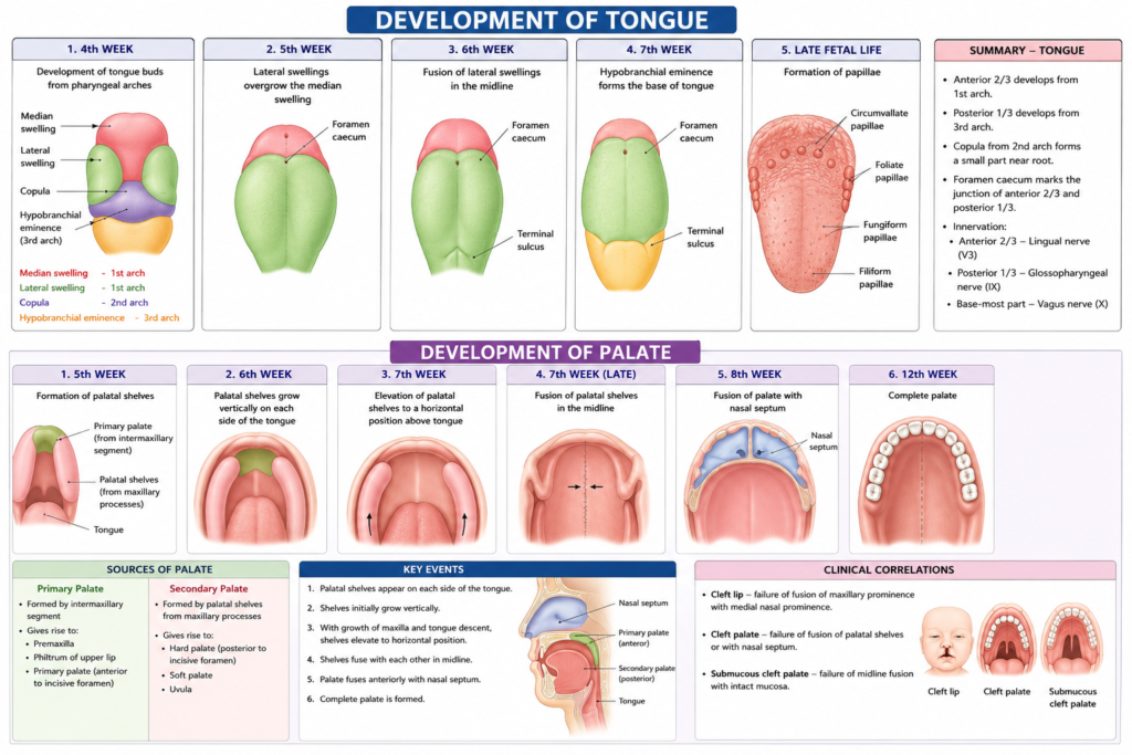
The tongue and palate develop alongside facial structures. Both regions hold special importance in oral biology.
Tongue Development
The tongue forms from multiple swellings. The anterior two-thirds arise from the first arch. The posterior one-third arises from the third arch. This dual origin explains the tongue’s different nerve supplies.
Palate Development
The palate forms in two stages:
- Primary palate – Forms from the frontonasal process
- Secondary palate – Forms from paired palatal shelves of maxillary processes
These shelves grow vertically first. Then, they rotate horizontally and fuse at the midline. Failure of this fusion causes cleft palate, a common congenital anomaly.
Applied Embryology in Dentistry
Clinical anomalies often trace back to embryonic development errors. Dentists encounter these conditions regularly in practice.
Cleft Lip and Palate
This anomaly results from failed fusion of facial processes. It affects feeding, speech, and dental alignment. Early surgical correction improves long-term outcomes significantly.
Branchial Cysts
These cysts develop from incomplete pharyngeal cleft closure. They appear as painless neck swellings. Surgical removal usually resolves this condition completely.
Mandibular Anomalies
Since the mandible originates from the first arch, disruptions here cause conditions like micrognathia. This condition leads to a smaller-than-normal lower jaw.
Tongue Anomalies
Failure of proper tongue fusion can cause ankyloglossia, commonly called tongue-tie. This condition restricts tongue movement and affects speech development.
Understanding the embryology of head, face, and neck helps dentists diagnose these conditions early. Early diagnosis, in turn, allows better treatment planning and outcomes.
Conclusion
The embryology of head, face, and neck explains how complex structures form from simple embryonic layers. Pharyngeal arches, facial processes, and palatal shelves all contribute to this remarkable transformation.
Dental professionals benefit greatly from this knowledge. It helps explain congenital anomalies like cleft palate, branchial cysts, and tongue-tie. Ultimately, understanding embryonic development supports better diagnosis and treatment across dental practice.
Frequently Asked Questions (FAQs)
Pharyngeal arches are paired embryonic structures that form the skeletal, muscular, nervous, and vascular framework of the head and neck region.
The first pharyngeal arch, also called the mandibular arch, forms the mandible and muscles of mastication.
Cleft lip and palate result from incomplete fusion of facial processes or palatal shelves during early embryonic development.
The tongue develops from multiple swellings, with the anterior portion arising from the first arch and the posterior portion from the third arch.
A branchial cyst forms due to incomplete closure of pharyngeal clefts, appearing as a painless swelling along the side of the neck.