The vertebral canal protects one of the most important structures in the human body: the spinal cord. Dental professionals often overlook this region, yet it plays a supporting role in local anesthesia, sedation, and pain pathways of the head and neck. This guide breaks down the contents of vertebral canal in plain language, so students, dentists, and curious readers can understand this anatomy without getting lost in jargon.
We will explore the spinal cord, spinal nerve roots, three protective membranes, cerebrospinal fluid, and the venous network that surrounds them. Along the way, you will find easy tables and a simple flowchart to visualize how each layer fits together.
What Is the Vertebral Canal?
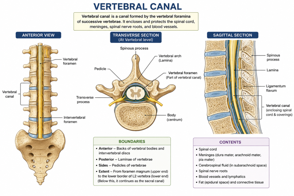
The vertebral canal is a long, bony tunnel formed by the vertebral foramina stacked one on top of another. It runs from the base of the skull down to the sacrum. This canal shields the spinal cord from injury while allowing nerves to branch out toward the rest of the body.
Think of it as a protective corridor. The walls are bone, the inner lining is soft tissue, and the core holds the spinal cord itself. Because the canal is narrow, any swelling or compression inside it can create serious problems. That is why understanding the contents of vertebral canal matters, especially for anyone working near the spine during medical or dental procedures.
Several structures sit inside this corridor. Each one has a distinct job, and together they keep the central nervous system safe and functional.
Contents of Vertebral Canal: Quick Overview
Before diving deeper, here is a simple table summarizing the main contents of vertebral canal.
| Structure | Location | Main Function |
|---|---|---|
| Spinal cord | Center of canal | Transmits nerve signals |
| Spinal nerve roots | Extend laterally | Carry motor and sensory information |
| Dura mater | Outermost meningeal layer | Tough protective covering |
| Arachnoid mater | Middle meningeal layer | Holds cerebrospinal fluid |
| Pia mater | Innermost meningeal layer | Clings directly to spinal cord |
| Cerebrospinal fluid | Subarachnoid space | Cushions and nourishes the cord |
| Internal vertebral venous plexus | Epidural space | Drains blood, connects to system veins |
| Fat and connective tissue | Epidural space | Fills empty spaces, absorbs shock |
This table gives a bird’s-eye view. Next, let’s look at each structure individually.
The Spinal Cord and Nerve Roots
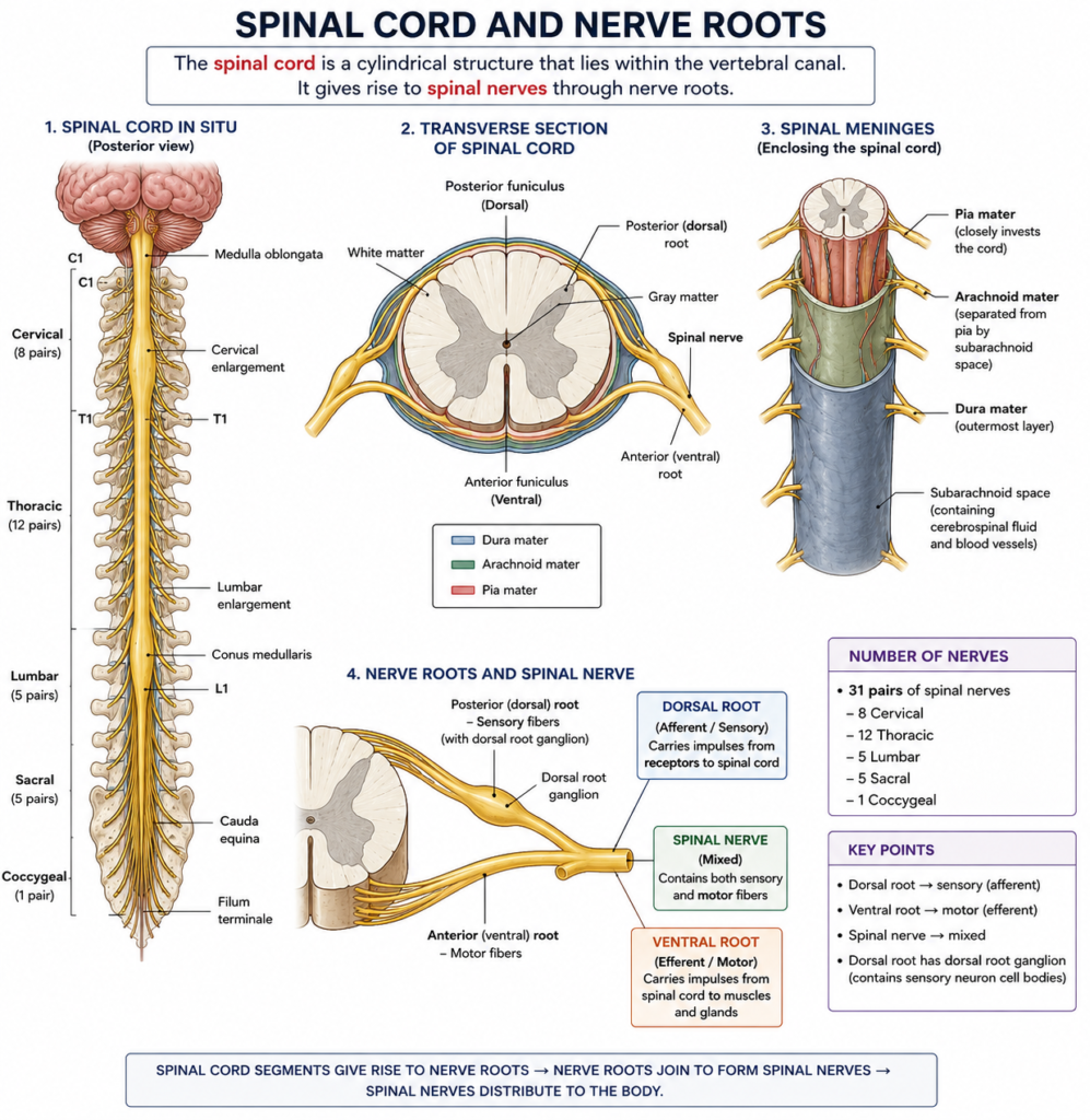
The spinal cord sits at the very center of the canal. It stretches from the medulla oblongata down to roughly the first or second lumbar vertebra in adults. Below that point, only nerve roots continue, forming a bundle called the cauda equina.
Thirty-one pairs of spinal nerves exit through small openings called intervertebral foramina. These nerves control movement, sensation, and reflexes throughout the body. Consequently, any pressure on the cord or its roots can cause pain, numbness, or weakness in distant areas, sometimes even the jaw or face region that dentists treat.
The cord itself is soft and gel-like. Therefore, it depends heavily on the surrounding membranes and fluid for protection.
The Three Meningeal Layers
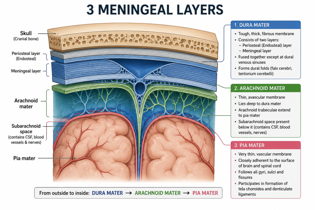
The meninges are three connective tissue layers wrapped around the spinal cord. Each layer contributes uniquely to the contents of vertebral canal, and dental students often study these layers alongside cranial anatomy since they connect directly with brain coverings.
Dura Mater
The dura mater forms the tough, outermost layer. It is thick, fibrous, and somewhat inflexible. This layer creates a loose-fitting tube around the spinal cord, leaving a gap called the epidural space between itself and the bony canal wall.
Anesthesiologists inject medication into this exact space during epidural procedures. As a result, understanding dura mater position is clinically valuable beyond just textbook knowledge.
Arachnoid Mater
Just beneath the dura lies the arachnoid mater, a delicate, web-like membrane. It sits close to the dura but is separated by a thin subdural space. Between the arachnoid mater and the pia mater lies the subarachnoid space, which holds cerebrospinal fluid.
This fluid acts like a shock absorber. It also helps remove waste products from nervous tissue. Meanwhile, the arachnoid mater keeps this fluid contained so it does not leak into surrounding tissues.
Pia Mater
The pia mater is the innermost and most delicate layer. It hugs the spinal cord tightly, following every contour and groove. Unlike the tougher dura mater, the pia mater is thin and highly vascular, supplying nutrients directly to the cord’s surface.
Small extensions of the pia mater, called denticulate ligaments, anchor the spinal cord to the dura mater on either side. This keeps the cord centered and stable inside the canal.
Flowchart: Layers From Outside to Inside
Here is a simple flowchart showing the order of structures, moving from the bony canal wall toward the spinal cord center.
Vertebral Bone (Canal Wall)
        ↓
Epidural Space (fat, connective tissue, system veins)
        ↓
Dura Mater
        ↓
Subdural Space
        ↓
Arachnoid Mater
        ↓
Subarachnoid Space (cerebrospinal fluid)
        ↓
Pia Mater
        ↓
Spinal Cord
This visual makes it easier to remember the sequence. First comes bone, then fat and veins, then three membranes, and finally the cord itself.
Cerebrospinal Fluid and Its Role
Cerebrospinal fluid, often shortened to CSF, fills the subarachnoid space. It circulates continuously, cushioning the spinal cord against sudden movements. Additionally, CSF helps regulate pressure inside the canal and removes metabolic waste.
Doctors sometimes sample this fluid through a lumbar puncture, inserted below the spinal cord’s end point to avoid injury. This procedure highlights why knowing the exact contents of vertebral canal, and their boundaries, is so important in clinical practice.
The Venous System Inside the Canal
Alongside the meninges, the epidural space contains a rich network of veins known as the internal vertebral venous plexus, or Batson’s plexus. This system veins network drains blood from the vertebrae, spinal cord, and surrounding tissues.
Unlike typical veins, this plexus has no valves. Therefore, blood can flow in multiple directions depending on pressure changes. This feature explains how infections or cancer cells sometimes spread between the pelvis, spine, and even skull base, since these veins connect widely throughout the body.
For dental professionals, this detail is fascinating because oral infections can theoretically travel through connected venous pathways under rare circumstances. While uncommon, it demonstrates how interconnected our body systems truly are.
Why This Anatomy Matters for Dental Practice
You might wonder why a dental website covers spinal anatomy at all. However, dentists frequently work with regional anesthesia, sedation dentistry, and pain referral patterns that originate from shared nerve pathways and connected vascular networks.
Understanding the broader nervous and venous systems helps dental professionals recognize unusual symptoms, communicate better with medical specialists, and appreciate how head, neck, and spinal structures relate. Moreover, many dental students study general human anatomy, including the vertebral canal, before specializing in oral and maxillofacial regions.
Summary Table: Meninges Comparison
| Layer | Texture | Position | Special Feature |
|---|---|---|---|
| Dura mater | Thick, fibrous | Outermost | Creates epidural space |
| Arachnoid mater | Thin, web-like | Middle | Holds CSF beneath it |
| Pia mater | Delicate, vascular | Innermost | Attached directly to cord |
Conclusion
The vertebral canal houses far more than empty space. From the spinal cord to the protective meninges, cerebrospinal fluid, and an extensive venous network, every structure plays a specific role in protecting our nervous system. Learning the contents of vertebral canal gives dental students and professionals a fuller picture of human anatomy, one that connects directly to sedation, pain patterns, and whole-body health. Next time you review head and neck anatomy, remember that the spine and skull are more connected than they first appear.
Frequently Asked Questions
The main contents of vertebral canal include the spinal cord, spinal nerve roots, three meningeal layers, cerebrospinal fluid, and the internal vertebral venous plexus within the epidural space.
Dura mater is the tough outer layer, arachnoid mater is the thin middle layer holding cerebrospinal fluid, and pia mater is the delicate inner layer attached directly to the spinal cord.
The epidural space contains fat, connective tissue, and system veins. It also serves as the injection site for epidural anesthesia during childbirth or certain surgeries.
No, the spinal cord ends around the first or second lumbar vertebra. Below this point, only nerve roots, called the cauda equina, continue through the canal.
Dentists study general anatomy, including the vertebral canal, to understand nerve pathways, sedation risks, and how infections could theoretically spread through connected vascular systems.