The human skull holds a hidden chamber that shields the brain from harm. This space plays a huge role in survival. It supports one of the most complex organs in the body. Below, we break down its structure, layers, and importance in simple terms.
What Is This Protective Skull Chamber?
This bony compartment sits inside the skull. It forms a hollow, dome-shaped space that surrounds and cushions the brain. Several skull bones fuse together to build its walls. As a result, the brain gets a strong, rigid shield against outside forces.
Inside this chamber, several tissues work together. These include the brain itself, protective membranes, fluid, nerves, and blood vessels. Therefore, this space is never truly empty. It stays busy supporting one of the body’s most important systems.
This structure belongs to a larger group of body spaces called the dorsal cavity. The spinal canal also falls under this group. Together, they protect the entire central nervous system, which controls almost everything the body does.
Bones That Form This Skull Space
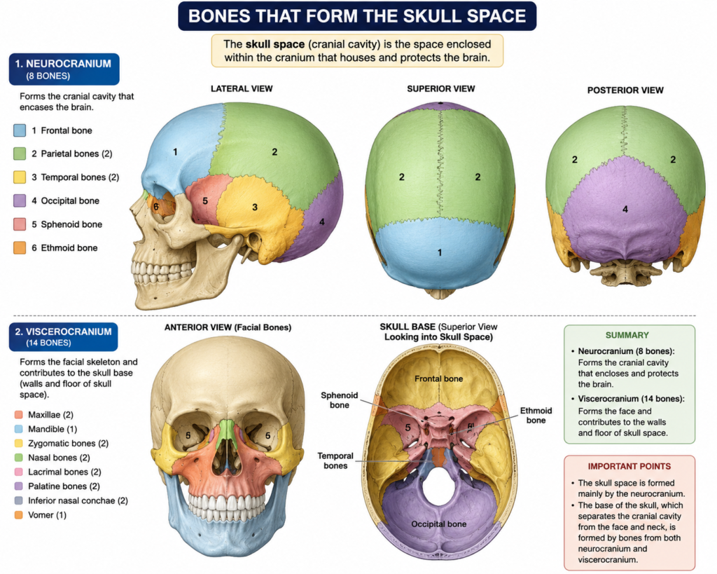
Several bones join together to create the walls of this chamber. Each one contributes a specific section. The table below breaks this down clearly.
| Bone | Location Formed |
|---|---|
| Frontal bone | Front wall |
| Parietal bones | Sides and roof |
| Temporal bones | Lower sides |
| Occipital bone | Back and base |
| Sphenoid bone | Floor and sides |
| Ethmoid bone | Front floor |
These bones meet at joints called sutures. In babies, these joints stay flexible. This flexibility allows the skull to pass through birth safely. Later, growth spurts also benefit from this flexibility. Eventually, the sutures harden and fuse permanently during childhood.
Three Regions Inside the Cranial Cavity
The cranial cavity divides into three separate zones. Each zone supports a different part of the brain. This layout helps distribute pressure evenly across the skull.
Front Zone (Anterior Fossa) This region sits above the eyes and nose. It supports the frontal lobes, which handle reasoning and decision-making.
Middle Zone (Middle Fossa) This area sits lower and more central. It holds the temporal lobes and the pituitary gland. Several nerve openings also pass through here.
Back Zone (Posterior Fossa) This is the largest and deepest region. It houses the cerebellum and brainstem. These structures manage balance, coordination, and breathing.
Layers That Protect the Brain
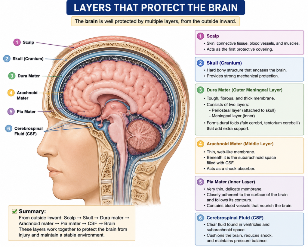
Multiple layers work together to guard the brain inside this space. First, three thin membranes called the meninges wrap around the brain tissue. Next, fluid fills the gaps between these membranes. Finally, bone forms the outer shield.
The flowchart below shows this protective order, moving from the brain outward.
Brain Tissue
     ↓
Pia Mater (innermost membrane)
     ↓
Subarachnoid Space (filled with fluid)
     ↓
Arachnoid Mater (middle membrane)
     ↓
Dura Mater (tough outer membrane)
     ↓
Skull Bone (final protective wall)
This layered system absorbs shock. It also reduces friction during movement. Meanwhile, cerebrospinal fluid circulates constantly, delivering nutrients and removing waste.
What Structures Live Inside This Space?
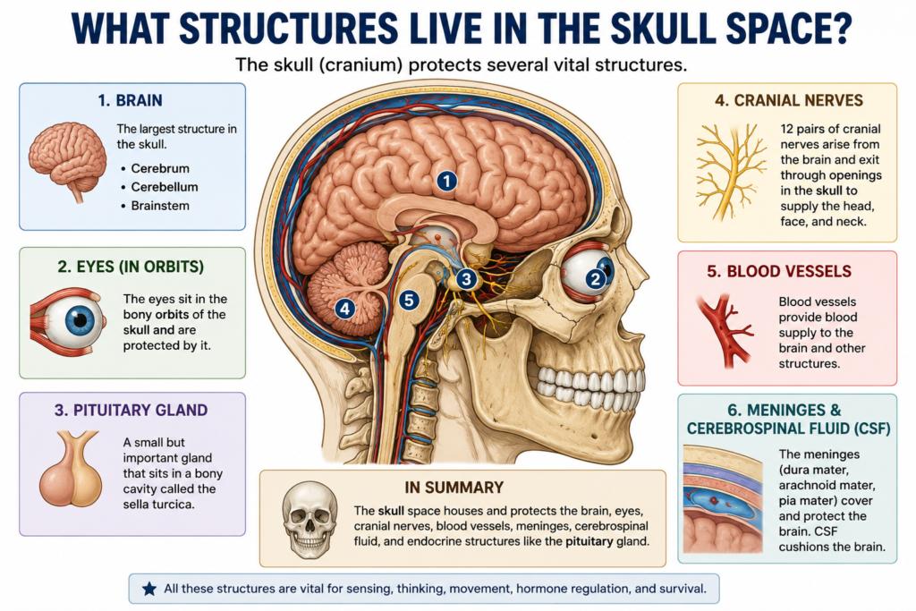
Many important parts exist within this region. Understanding each one helps explain why this space matters so much.
- Brain tissue, including the cerebrum, cerebellum, and brainstem
- Twelve pairs of cranial nerves
- Arteries and veins supplying blood flow
- Protective membranes, known as the meninges
- Cerebrospinal fluid, which cushions and nourishes the brain
Each part depends on the others. For instance, blood vessels feed oxygen to nerve tissue. Meanwhile, fluid keeps pressure levels balanced. Without this teamwork, brain function would suffer quickly.
Why This Structure Matters So Much
The brain controls nearly every action the body performs. Therefore, protecting it becomes a top priority for the skeletal system. Bone alone cannot absorb every impact. That is why fluid and membranes add extra cushioning.
This chamber also stays completely enclosed. Consequently, any swelling or bleeding inside raises pressure fast. Doctors call this rise in pressure “intracranial pressure.” It can become dangerous within a short time.
Because of this risk, head injuries always receive urgent medical attention. Even a minor impact can cause internal swelling. As a result, monitoring pressure levels becomes critical during treatment.
Common Conditions Linked to This Region
Several medical conditions relate directly to this skull space. Recognizing them helps explain why anatomy knowledge matters in real medical settings.
- Traumatic brain injury from falls or accidents
- Bleeding inside the skull, known as hemorrhage
- Fluid buildup, called hydrocephalus
- Skull fractures from strong impacts
- Pressure increases caused by tumors or swelling
Doctors often use CT scans or MRI machines to check for these issues. These tools reveal swelling, bleeding, or structural damage quickly. Early detection often improves recovery outcomes significantly.
How This Space Differs From Other Body Cavities
The body contains several major cavities. Each one protects different organs. For example, the thoracic cavity protects the heart and lungs. Similarly, the abdominal cavity protects digestive organs.
This skull chamber stands apart, though. It houses the control center for the entire nervous system. No other cavity carries such critical responsibility for whole-body function.
Conclusion
This hidden skull chamber does far more than hold the brain in place. It works as a full protection system, combining bone, fluid, and membrane layers. Every piece plays a role in keeping the brain safe during daily movement and unexpected impacts.
Learning how this space works helps explain why head injuries deserve fast medical care. It also shows how beautifully the human body defends its most valuable organ. Anyone studying anatomy, medicine, or general health can benefit from understanding this remarkable structure.
Frequently Asked Questions
1. What is the main job of the cranial cavity? Its main job involves protecting the brain from injury and supporting its structure within the skull.
Its main job involves protecting the brain from injury and supporting its structure within the skull.
The frontal, parietal, temporal, occipital, sphenoid, and ethmoid bones all contribute to its walls.
Cerebrospinal fluid cushions the brain and removes waste products continuously.
Yes, swelling raises internal pressure quickly since the space cannot expand, which can harm brain tissue.
Doctors typically use CT scans or MRI scans to detect bleeding, swelling, or structural damage.