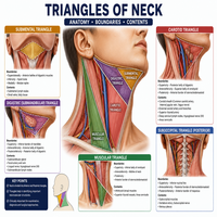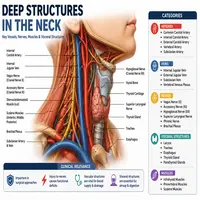
Deep Structures in the Neck: A Complete Anatomy Guide
The neck is small. Yet it holds some of the busiest anatomy in the human body. Nerves, blood vessels, muscles, and glands all pack into this narrow space. Understanding the deep structures in the neck matters for students, clinicians, and anyone curious about human anatomy.
This guide breaks down the neck layer by layer. We will cover the fascia, muscles, vessels, nerves, and visceral organs. Additionally, we will explain why these structures matter clinically. So, let’s dive in.
Why the Neck’s Anatomy Is So Complex
The neck connects the head to the trunk. Therefore, it must carry every major nerve, vessel, and airway between these two regions. This creates a crowded anatomical space.
Furthermore, the neck supports movement. Muscles here allow rotation, flexion, and extension. Meanwhile, deeper layers protect vital organs like the trachea and esophagus.
Because of this density, surgeons and radiologists study the neck in layers. Each layer has a name, a boundary, and a function. Let’s start with the fascia, since it organizes everything else.
The Fascial Layers of the Neck
Fascia acts like packaging material. It wraps muscles, vessels, and organs into organized compartments. Consequently, understanding fascia helps explain how infections or fluids spread through the neck.
There are three main fascial layers:
| Fascial Layer | Location | Structures Enclosed |
|---|---|---|
| Superficial (investing) layer | Just under the skin | Sternocleidomastoid, trapezius |
| Pretracheal layer | Anterior neck | Thyroid gland, trachea, esophagus |
| Prevertebral layer | Posterior neck | Vertebral column, deep muscles |
Between these layers, potential spaces exist. For instance, the retropharyngeal space allows infections to travel from the throat down toward the chest. This is why dentists and ENT specialists pay close attention to fascial planes.
Deep Muscles in the Neck
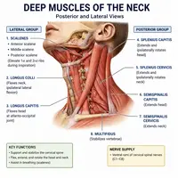
Now let’s move to the muscles. The deep structures in the neck include several muscle groups that most people never think about.
These muscles fall into a few categories:
- Prevertebral muscles – longus colli, longus capitis
- Scalene muscles – anterior, middle, and posterior scalenes
- Suboccipital muscles – control fine head movements
The scalene muscles deserve special mention. They attach to the cervical vertebrae and first two ribs. Between the anterior and middle scalenes lies a narrow gap called the scalene triangle. Notably, the brachial plexus and subclavian artery pass through this exact space.
As a result, tightness in these muscles can compress nerves. This condition is known as thoracic outlet syndrome. So, these small muscles carry big clinical weight.
Major Vessels Hidden Beneath the Surface
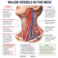
Blood vessels form another critical part of the deep structures in the neck. The carotid sheath houses three key structures:
- Common carotid artery
- Internal jugular vein
- Vagus nerve
This sheath runs vertically along both sides of the neck. Meanwhile, smaller vessels branch off to supply the thyroid, larynx, and facial structures.
Additionally, the vertebral arteries travel through openings in the cervical vertebrae. They supply blood to the brainstem and cerebellum. Because these arteries sit so close to bone, neck injuries can sometimes affect blood flow to the brain.
Nerves That Run Through the Neck
Nerves crisscross the neck in a complex pattern. The cervical plexus and brachial plexus both originate here. Meanwhile, several cranial nerves pass through on their way to the chest or face.
Here is a simple flow of nerve pathways through the neck:
Brainstem
   │
   ▼
Cranial Nerves (IX, X, XI, XII) exit skull base
   │
   ▼
Pass through carotid sheath region
   │
   ▼
Branch to larynx, pharynx, shoulder, and tongue muscles
The vagus nerve (cranial nerve X) deserves attention. It travels the entire length of the neck inside the carotid sheath. Along the way, it sends branches to the larynx, controlling voice production. Consequently, neck surgery near this area carries a risk of voice changes.
The phrenic nerve also passes through the neck. It arises from cervical roots C3 to C5 and travels down to the diaphragm. Interestingly, this means the diaphragm’s nerve supply originates far from the chest.
Visceral Structures: Trachea, Esophagus, and Thyroid
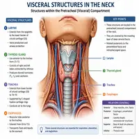
Beyond muscles and vessels, the neck contains vital visceral organs. These sit within the pretracheal fascia layer.
| Organ | Function | Clinical Note |
|---|---|---|
| Trachea | Air passage to lungs | Can be compressed by thyroid enlargement |
| Esophagus | Food passage to stomach | Sits just behind the trachea |
| Thyroid gland | Hormone regulation | Wraps around the trachea’s sides |
| Parathyroid glands | Calcium regulation | Tiny glands behind the thyroid |
Notably, the thyroid gland sits very close to the recurrent laryngeal nerves. Surgeons must identify these nerves carefully during thyroid operations. Otherwise, voice complications can occur.
Clinical Relevance of Deep Neck Anatomy
Why does all this matter beyond the classroom? Because the deep structures in the neck are frequently involved in real medical scenarios.
For example:
- Deep neck infections can spread along fascial planes toward the mediastinum.
- Thyroid surgery requires precise knowledge of nerve and vessel locations.
- Trauma cases often involve the carotid sheath, risking major bleeding.
- Nerve blocks for shoulder surgery target the brachial plexus in the neck.
Therefore, radiologists, surgeons, and emergency physicians rely heavily on this anatomical knowledge daily. A small misstep in this region can lead to significant complications.
A Quick Summary Table
| Layer/System | Key Structures | Main Function |
|---|---|---|
| Fascia | Superficial, pretracheal, prevertebral | Organizes and protects |
| Muscles | Scalenes, prevertebral group | Movement and support |
| Vessels | Carotid sheath, vertebral arteries | Blood supply |
| Nerves | Vagus, phrenic, cervical plexus | Motor and sensory control |
| Viscera | Trachea, esophagus, thyroid | Breathing, digestion, hormones |
Conclusion
The neck may look simple from the outside. However, it hides an intricate network of muscles, vessels, nerves, and organs. Learning the deep structures in the neck helps explain many clinical conditions, from voice changes after surgery to nerve compression syndromes.
Moreover, this knowledge is not just for medical students. Physical therapists, dentists, and even fitness professionals benefit from understanding how these layers interact. Ultimately, a clear picture of neck anatomy leads to safer treatments and better patient outcomes.
Keep this guide handy as a quick reference. Anatomy becomes much easier once you see how each layer connects to the next.
Frequently Asked Questions
They include the prevertebral muscles, carotid sheath vessels, vagus and phrenic nerves, cervical vertebrae, and visceral organs like the trachea and esophagus, all wrapped in deep fascial layers.
The carotid sheath protects three vital structures: the common carotid artery, internal jugular vein, and vagus nerve. Damage here can cause serious bleeding or nerve dysfunction.
The scalene muscles, longus colli, longus capitis, and suboccipital muscles are classified as deep neck muscles. They support posture and fine head movements.
The thyroid sits close to the recurrent laryngeal nerves and parathyroid glands. Surgeons must navigate these deep structures carefully to avoid voice or calcium regulation problems.
Yes. Fascial planes, like the retropharyngeal space, can allow infections to travel from the throat down toward the chest, making deep neck infections potentially dangerous.