Hemostasis: How the Body Stops Bleeding Naturally
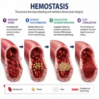
Blood loss can be dangerous. Fortunately, the body has a built-in defense system. This system is called hemostasis. It stops bleeding quickly and efficiently after an injury.
Hemostasis involves several coordinated steps. Blood vessels, platelets, and clotting proteins all work together. Meanwhile, the body also controls this process carefully to avoid unwanted clots. Let’s explore how this remarkable system functions.
What Is Hemostasis?
Hemostasis literally means “stopping blood.” It refers to the process that prevents excessive bleeding when a vessel is damaged. Without it, even a small cut could become life-threatening.
The process happens in stages. First, the vessel constricts. Then, platelets gather at the injury site. Finally, a stable clot forms through a chemical cascade. Each stage builds on the previous one.
Interestingly, hemostasis must also stay balanced. Too much clotting can block healthy vessels. Too little can cause dangerous bleeding. Therefore, the body regulates this system with precision.
The Three Main Stages of Hemostasis
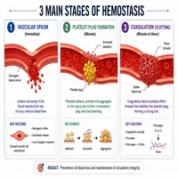
Hemostasis generally unfolds in three connected phases. Understanding each phase helps clarify how the whole process works.
| Stage | Main Event | Duration |
|---|---|---|
| Vascular spasm | Blood vessel constricts | Seconds |
| Platelet plug formation | Platelets stick together | Minutes |
| Coagulation | Fibrin mesh stabilizes the clot | Minutes to hours |
These three stages overlap significantly. As a result, the body doesn’t wait for one phase to finish before starting the next. This overlapping design speeds up the response to injury.
Stage One: Vascular Spasm
The first response to injury is vasoconstriction. Smooth muscle in the vessel wall contracts immediately. Consequently, blood flow slows down at the injury site.
This reaction happens through local nerve reflexes and chemical signals. Endothelin, a protein released by damaged vessel walls, plays a major role here. Additionally, pain receptors trigger nervous system responses that support constriction.
This step is brief but important. It buys time for the next stages to begin. Without this initial constriction, blood loss would be much harder to control.
Stage Two: Platelet Plug Formation
Next, platelets take center stage. When a vessel is damaged, collagen fibers become exposed. Platelets then stick to this collagen, a process called adhesion.
Once attached, platelets become activated. They change shape and release chemical signals. These signals attract more platelets to the area. This is known as platelet aggregation.
Here is a simple flow of this stage:
Vessel Injury
     │
     ▼
Collagen Exposed
     │
     ▼
Platelets Adhere (via von Willebrand factor)
     │
     ▼
Platelets Activate and Release Signals
     │
     ▼
More Platelets Aggregate → Platelet Plug Formed
This plug is fragile at first. Therefore, it needs reinforcement. That reinforcement comes from the coagulation cascade, which we’ll cover next.
Stage Three: Coagulation Cascade
Coagulation strengthens the platelet plug. It transforms a loose cluster of platelets into a stable, durable clot. This process relies on clotting factors, which are proteins found in the blood plasma.
There are two pathways that begin this cascade:
- Intrinsic pathway – triggered by damage inside the vessel
- Extrinsic pathway – triggered by damage outside the vessel, releasing tissue factor
Both pathways eventually merge into a common pathway. This shared pathway converts prothrombin into thrombin. Thrombin then converts fibrinogen into fibrin. Fibrin strands weave through the platelet plug, creating a mesh-like net.
This mesh traps blood cells and hardens the clot. Consequently, the wound becomes sealed and protected while healing begins underneath.
Key Clotting Factors Involved
Several clotting factors participate in this cascade. Each one has a specific numbered designation.
| Factor Number | Common Name | Role |
|---|---|---|
| I | Fibrinogen | Converted to fibrin |
| II | Prothrombin | Converted to thrombin |
| III | Tissue factor | Starts extrinsic pathway |
| IV | Calcium ions | Required for multiple steps |
| VIII | Antihemophilic factor | Deficiency causes hemophilia A |
Notably, vitamin K plays a supporting role. It helps the liver produce several clotting factors. Without enough vitamin K, clotting ability weakens significantly.
How the Body Prevents Excess Clotting
Hemostasis must stop once the wound heals. Otherwise, clots could block healthy blood vessels. Therefore, the body has natural anticoagulant mechanisms.
Antithrombin, for example, inactivates thrombin once it’s no longer needed. Meanwhile, protein C and protein S break down certain clotting factors. Additionally, the fibrinolytic system dissolves clots gradually using an enzyme called plasmin.
This balance is essential for health. Too much clotting can lead to conditions like deep vein thrombosis. Too little clotting can lead to bleeding disorders. As a result, hemostasis represents a finely tuned biological balance.
Clinical Relevance of Hemostasis
Understanding hemostasis matters far beyond textbooks. Many medical conditions relate directly to this process.
For instance:
- Hemophilia results from missing clotting factors.
- Thrombosis occurs when clots form unnecessarily inside vessels.
- Anticoagulant medications like warfarin interfere with clotting factor production.
- Vitamin K deficiency can impair normal clot formation.
Doctors often test hemostasis using blood panels. These tests measure clotting time and factor levels. Consequently, this physiology directly guides treatment decisions in hospitals every day.
Quick Summary Table
| Stage | Main Players | Purpose |
|---|---|---|
| Vascular spasm | Smooth muscle, endothelin | Reduce blood flow immediately |
| Platelet plug | Platelets, von Willebrand factor | Temporary seal |
| Coagulation | Clotting factors, fibrin | Stable, durable clot |
| Clot breakdown | Plasmin, fibrinolysis | Remove clot after healing |
Conclusion
Hemostasis is one of the body’s most elegant survival systems. It moves through three connected stages, each building on the last. From vascular spasm to platelet plugs to coagulation, this process protects the body from dangerous blood loss.
Moreover, hemostasis doesn’t work alone. Anticoagulant systems keep it in check, preventing harmful clot formation. This balance between clotting and anti-clotting keeps the circulatory system healthy.
Understanding hemostasis helps explain many medical conditions, from hemophilia to thrombosis. Whether you’re a student or simply curious about the body, this knowledge reveals just how well-designed human physiology truly is.
Frequently Asked Questions
Hemostasis is the body’s natural process for stopping bleeding after a blood vessel is injured. It involves vessel constriction, platelet clumping, and clot formation.
The three stages are vascular spasm, platelet plug formation, and coagulation. Together, they stop bleeding and stabilize the wound site.
Platelets stick to exposed collagen at an injury site. They then release signals that attract more platelets, forming a temporary plug over the wound.
Vitamin K helps the liver produce several essential clotting factors. Without adequate vitamin K, the coagulation cascade becomes impaired.
Malfunctions can lead to either excessive bleeding, as seen in hemophilia, or unwanted clot formation, as seen in thrombosis and deep vein thrombosis.