Cell Structure and Function
Every living organism, whether a single bacterium or a complex human body, is built from a basic unit called the cell. This microscopic structure carries out all the chemical and physical activities needed for life, and therefore it is often described as the smallest functional unit of living matter. In physiology, understanding cell is essential, because every tissue, organ, and system ultimately depends on how individual units behave, communicate, and reproduce.
Cells vary enormously in shape and size, ranging from tiny bacteria to large nerve fibers. However, despite this diversity, most units share a common architecture: an outer boundary, an internal fluid matrix, and a set of specialized internal structures. In the sections that follow, this chapter explores each of these components in detail, moving from the outer boundary inward to the nucleus, and finally to the process of programmed death.
Structure of the Cell
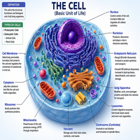
A typical animal unit is organized into three broad regions. First, there is the outer boundary, which separates the inside from the outside environment. Second, there is the fluid-filled interior, which houses dissolved substances and small structures. Third, there is a centrally located control center that stores genetic material.
Although the internal arrangement of cell is consistently organized, the overall shape often reflects function. For instance, a nerve fiber is long and thread-like, allowing it to transmit signals over distance, while a red blood corpuscle is flattened and disc-shaped, allowing efficient gas exchange. Consequently, structure and function are tightly linked at every level of biological organization.
The table below summarizes the major structural regions and their general roles.
| Structural Region | General Description | Primary Role |
|---|---|---|
| Outer boundary | Thin, flexible lipid layer | Regulates entry and exit of substances |
| Fluid interior | Gel-like substance filling the interior | Houses organelles and metabolic reactions |
| Internal organelles | Specialized membrane and non-membrane structures | Carry out specific metabolic tasks |
| Control center | Dense structure containing genetic material | Directs growth, repair, and division |
The Cell Membrane
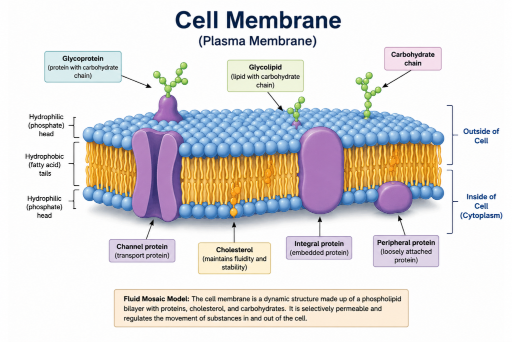
The outer boundary, commonly called the plasma membrane, is composed mainly of a phospholipid bilayer embedded with proteins, cholesterol, and carbohydrate chains. This arrangement is frequently described using the fluid mosaic model, since the lipid molecules move freely while proteins float within the layer like icebergs in a sea.
Functionally, this boundary is selectively permeable. As a result, small, non-polar molecules such as oxygen and carbon dioxide pass through with ease, whereas larger or charged particles require specific transport proteins. Several transport mechanisms are involved, including diffusion, osmosis, facilitated transport, and active transport driven by energy.
In addition to transport, the boundary plays a vital role in communication. Surface receptors detect hormones and signaling molecules, and signals are subsequently relayed into the interior. Without this communication system, coordinated physiological responses, such as the release of insulin in reaction to glucose, would not be possible.
Cytoplasm: The Cellular Matrix
Beneath the outer boundary lies the cytoplasm, a semi-fluid substance that fills the entire interior space. It consists of cytosol, which is mostly water mixed with ions, proteins, and nutrients, along with the various internal structures suspended within it.
Metabolically, the cytoplasm is far from passive. Numerous enzymatic reactions, including glycolysis, occur directly within this matrix. Moreover, it provides a medium through which nutrients, waste products, and signaling molecules diffuse between internal structures. Therefore, the cytoplasm functions as both a physical support and a busy metabolic workspace.
Organelles in the Cytoplasm
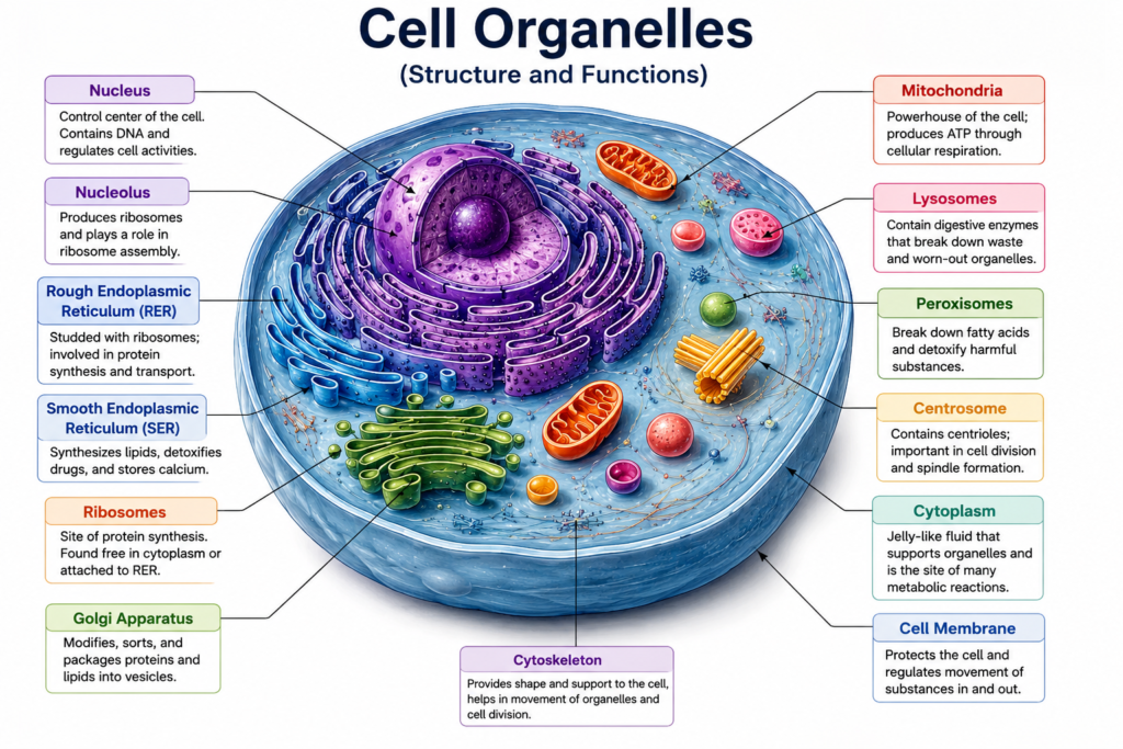
Suspended within the cytoplasm are numerous specialized structures known as organelles, each performing a distinct task. Broadly, these structures are classified into two categories based on whether they possess a limiting membrane. This classification is useful because membrane-bound structures can maintain a separate internal environment, while non-membrane structures cannot.
The flowchart below illustrates this classification.
                     Organelles
                         |
        -----------------------------------
        |                                  |
Membrane-Bound Organelles        Non-Membrane-Bound Organelles
        |                                  |
 Mitochondria                        Ribosomes
 Endoplasmic Reticulum               Centrioles
 Golgi Apparatus                     Cytoskeleton
 Lysosomes
 Peroxisomes
Organelles With a Limiting Membrane
Membrane-bound structures are enclosed by their own lipid layer, which allows them to maintain a chemical environment distinct from the surrounding cytoplasm. As a consequence, these structures can carry out specialized, often sensitive, biochemical reactions safely.
| Organelle | Membrane Type | Main Function |
|---|---|---|
| Mitochondria | Double membrane | Produces energy through respiration |
| Endoplasmic reticulum | Single membrane network | Synthesizes proteins and lipids |
| Golgi apparatus | Stack of single membranes | Packages and modifies secretory products |
| Lysosomes | Single membrane | Digests waste and damaged material |
| Peroxisomes | Single membrane | Breaks down fatty acids and toxins |
Mitochondria, for example, are often called the powerhouse, since they generate most of the chemical energy required for activity. Meanwhile, lysosomes act as a recycling center, breaking down worn-out components so that raw materials can be reused. Thus, each membrane-bound structure contributes a unique service to overall function.
Organelles Without a Limiting Membrane
In contrast, some structures lack a surrounding lipid layer altogether. Although they are not isolated chemically, these structures remain essential to survival and division.
| Organelle | Membrane Status | Main Function |
|---|---|---|
| Ribosomes | No membrane | Synthesizes proteins from genetic instructions |
| Centrioles | No membrane | Organizes spindle fibers during division |
| Cytoskeleton | No membrane | Provides shape, support, and movement |
Ribosomes, in particular, are scattered throughout the cytoplasm and are also attached to the endoplasmic reticulum, where they translate genetic code into functional proteins. Similarly, centrioles become especially active during division, ensuring chromosomes are distributed accurately to daughter structures.
Nucleus and Programmed Death
At the center of most units lies the nucleus, a membrane-bound control structure containing chromatin, nucleoli, and genetic material. This control center directs growth, metabolism, and reproduction by regulating which genes are expressed at any given time. Consequently, damage to the nucleus often has severe consequences for survival.
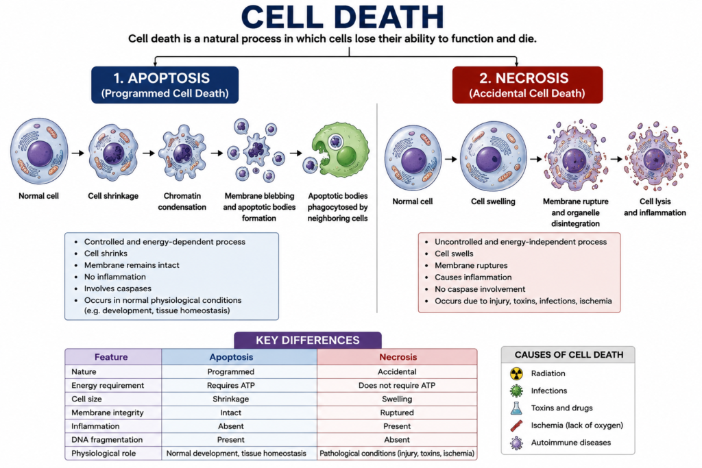
When a unit is irreparably damaged, aged, or no longer needed, it undergoes a regulated process of death. Two major pathways exist: necrosis, which is typically accidental and inflammatory, and apoptosis, which is a controlled, programmed process that avoids damaging surrounding tissue. The sequence below outlines programmed death.
Damage or Signal Detected
        |
Internal Checkpoint Activated
        |
Enzymes (Caspases) Triggered
        |
DNA Fragmentation Occurs
        |
Structure Shrinks and Breaks Apart
        |
Debris Cleared by Phagocytic Cells
This orderly removal process is essential for development, immune regulation, and prevention of cancer. Without it, damaged or excess units would accumulate, ultimately disrupting normal physiology.
Conclusion
In summary, the cell stands as the fundamental building block of physiology, organized into an outer boundary, a metabolically active cytoplasm, and a variety of organelles, both membrane-bound and non-membrane-bound. Each component, from the selectively permeable membrane to the regulatory nucleus, works together to sustain life. Furthermore, the orderly process of programmed death ensures that damaged or unnecessary structures are removed without harming surrounding tissue. Altogether, this coordinated organization explains why even the smallest unit of life can support something as complex as a fully functioning organism.
Frequently Asked Question
The basic unit of life is referred to as the cell, since it carries out every essential function required for survival.
Membrane-bound structures are enclosed by their own lipid layer and maintain a separate internal environment, whereas non-membrane structures lack this enclosure and function directly within the surrounding cytoplasm.
It is considered the control center because it stores genetic material and regulates which instructions are carried out at any given time.
Programmed death is typically triggered by internal damage, developmental signals, or external instructions that activate specific enzymes responsible for controlled breakdown.
Surface receptors located on the membrane detect external signals, such as hormones, and relay this information inward, allowing a coordinated physiological response.