Classification of Muscles: Types, Structure & Function
Muscles power almost everything the body does. From blinking to running a marathon, every movement depends on muscle tissue working correctly. Understanding the classification of muscles helps students, healthcare workers, and fitness enthusiasts grasp how the body moves, breathes, and pumps blood.
In this guide, we will break down muscle types by structure, control, and location. We will also explore real examples, a comparison table, a simple flowchart, and answers to common questions to make the concept easy to remember.
What Is Muscle Classification?
The classification of muscles refers to grouping muscle tissue based on shared features. Scientists typically classify muscles using two main criteria: structure and control.
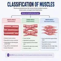
Structurally, muscles fall into three categories: skeletal, smooth, and cardiac. Meanwhile, based on control, muscles are either voluntary or involuntary. Therefore, a single muscle type often fits into both categories at once. For instance, skeletal muscle is voluntary, whereas smooth and cardiac muscles are involuntary.
Additionally, muscles differ in appearance under a microscope. Some appear striped, or “striated,” while others look smooth and uniform. This visual difference gives researchers another clue for accurate classification. Consequently, students often learn to identify muscle type simply by studying a tissue sample.
Classification of Muscles Based on Structure
Structure-based classification is the most common method taught in physiology. Below is a quick comparison table to simplify the differences.
| Muscle Type | Appearance | Location | Control | Nuclei per Cell |
|---|---|---|---|---|
| Skeletal Muscle | Striated | Attached to bones | Voluntary | Multiple |
| Smooth Muscle | Non-striated | Walls of internal organs | Involuntary | Single |
| Cardiac Muscle | Striated | Heart wall only | Involuntary | Single or double |
As shown above, striation is not exclusive to voluntary muscle. Cardiac muscle is striated too, yet it works involuntarily. This distinction often confuses beginners, so keep the table handy for quick reference.
Skeletal Muscle: The Movement Machine
Skeletal muscle attaches to bones through tendons. Consequently, it enables walking, lifting, chewing, and countless other movements. Under a microscope, this tissue shows visible stripes, known as striations, caused by organized protein filaments.
Furthermore, skeletal muscle fibers are long, cylindrical, and multinucleated. Since these muscles respond to conscious commands from the brain, they are labeled voluntary. However, some reflex actions bypass conscious thought entirely, even though they still use skeletal muscle.
Common examples include the biceps, quadriceps, and abdominal muscles. Athletes, physiotherapists, and trainers focus heavily on skeletal muscle because it directly affects strength, posture, and mobility. Regular exercise strengthens this tissue, while inactivity weakens it over time.
Smooth Muscle: The Silent Worker
Unlike skeletal muscle, smooth muscle lacks striations. It appears spindle-shaped and contains a single nucleus per cell. This muscle type lines hollow organs such as the stomach, intestines, blood vessels, and bladder.
Smooth muscle operates involuntarily. In other words, you cannot consciously control digestion or blood vessel constriction. Instead, the autonomic nervous system and hormones regulate its activity automatically.
Moreover, smooth muscle contracts slowly but sustains contractions longer than skeletal muscle. This feature suits its role in processes like peristalsis, where food moves gradually through the digestive tract. Meanwhile, blood vessels rely on smooth muscle to widen or narrow, controlling blood pressure throughout the day.
Cardiac Muscle: The Heart’s Engine
Cardiac muscle exists only in the heart. Like skeletal muscle, it shows striations under a microscope. Yet, similar to smooth muscle, it functions involuntarily and never tires throughout a person’s lifetime.
Cardiac cells connect through structures called intercalated discs. These discs allow electrical signals to pass quickly between cells, letting the heart contract as one coordinated unit. As a result, the heart beats rhythmically without conscious effort.
Interestingly, cardiac muscle combines features from both other muscle types. It borrows the striated look from skeletal muscle and the involuntary control from smooth muscle. This unique blend makes cardiac tissue a favorite topic in physiology exams and a critical focus in cardiology.
Classification of Muscles Based on Control
Beyond structure, classifying muscles based on control offers another useful lens. Voluntary muscles, namely skeletal muscles, respond to deliberate signals from the brain. On the other hand, involuntary muscles, including smooth and cardiac types, function automatically.
This distinction matters clinically too. For example, doctors assess voluntary muscle strength during physical exams, while involuntary muscle function often requires specialized tests like ECGs or endoscopies. Therefore, both categories play a distinct diagnostic role in modern medicine.
How Muscles Are Named
Anatomists often name muscles based on additional clues, such as size, shape, location, or number of origins. For example, “biceps” indicates two points of origin, while “deltoid” refers to a triangular shape.
Similarly, some muscles are named after their action, like the flexor or extensor muscles. Others take names from their location, such as the intercostal muscles found between the ribs. This naming system, though separate from structural classification, often helps students remember muscle function faster.
A Simple Flowchart for Quick Revision
                 MUSCLE TISSUE
                       |
        -------------------------------
        |                             |
   STRUCTURE-BASED               CONTROL-BASED
        |                             |
  -------------             -------------------
  |     |     |             |                 |
Skeletal Smooth Cardiac   Voluntary       Involuntary
(Striated)(Non-  (Striated) (Skeletal)   (Smooth + Cardiac)
         striated)
This flowchart offers a fast visual summary. Whenever you need a quick revision before an exam, this diagram works better than memorizing paragraphs.
Why Muscle Classification Matters
Understanding muscle classification isn’t just academic. Physiotherapists design rehabilitation plans around skeletal muscle recovery. Cardiologists monitor cardiac muscle health to prevent heart disease. Meanwhile, gastroenterologists study smooth muscle disorders like irritable bowel syndrome.
Additionally, fitness trainers use this knowledge to design workouts that target specific voluntary muscle groups. Without a clear grasp of classification, diagnosing or training the body accurately becomes far more difficult.
Conclusion
To sum up, the classification of muscles into skeletal, smooth, and cardiac types gives us a clear roadmap for understanding human movement and internal function. Skeletal muscle handles voluntary movement, smooth muscle manages internal organ activity, and cardiac muscle keeps the heart beating tirelessly.
Whether you’re a student preparing for exams, a healthcare professional, or simply curious about your own body, mastering this classification builds a strong foundation in physiology. Keep the table, flowchart, and FAQs below as quick references, and you’ll never confuse these muscle types again.
Frequently Asked Questions
The three main types are skeletal, smooth, and cardiac muscle. Each type differs in structure, location, and control.
Skeletal muscle is voluntary. It responds to conscious commands from the brain, allowing deliberate movement.
Cardiac muscle shows striations like skeletal muscle, yet it functions involuntarily like smooth muscle. This rare combination sets it apart from other tissue types.
Smooth muscle lines hollow internal organs, including the stomach, intestines, bladder, and blood vessels. It supports digestion, circulation, and excretion.
Classification of muscles guides diagnosis and treatment. Doctors, physiotherapists, and trainers use this knowledge to address issues specific to voluntary or involuntary muscle groups.