Pathology of Pulp and Periapex: How Dental Infections Progress
A toothache rarely appears without warning. Behind most cases of severe dental pain lies a chain of biological events that begins deep inside the tooth and, if untreated, spreads into the surrounding bone. Endodontists study this chain closely because understanding it allows them to intervene before irreversible damage occurs.
This article walks through the pathology of pulp and periapex, explaining how healthy tissue becomes inflamed, infected, and eventually destroyed. Along the way, you will find tables, a flowchart, and a concept map that break down each stage clearly. By the end, you will understand why early diagnosis matters so much in endodontic care.
Understanding Pulp and Periapical Anatomy
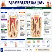
The dental pulp sits at the center of the tooth, surrounded by dentin and enamel, and contains blood vessels, nerves, and connective tissue. This soft tissue nourishes the tooth from within and provides sensory feedback when stimulated by heat, cold, or pressure. Just beyond the root tip lies the periapex, the region of bone and ligament surrounding the root apex.
These two areas function as a connected system rather than isolated structures. Consequently, disease rarely stays confined to one location for long. When the pulp becomes inflamed or infected, bacteria and their byproducts eventually travel through the root canal system toward the periapex, triggering a secondary inflammatory response in the surrounding bone.
Because this relationship is so direct, the pathology of pulp and periapex is often studied as a single progressive disease process rather than two separate conditions. Recognizing this connection helps clinicians predict how untreated pulp disease will likely evolve over time.
Causes of Pulp Pathology
Several factors can trigger inflammation or infection within the pulp, though bacterial invasion remains the most common cause. The table below summarizes the primary contributors.
| Cause | Mechanism | Typical Source |
|---|---|---|
| Dental caries | Bacteria penetrate dentin and reach the pulp | Untreated tooth decay |
| Trauma | Physical injury disrupts blood supply | Falls, sports injuries, accidents |
| Iatrogenic damage | Heat or mechanical injury during dental procedures | Deep cavity preparation, crown placement |
| Chemical irritation | Toxic materials placed near the pulp | Certain restorative or bleaching agents |
| Cracked tooth | Fracture allows bacterial entry | Chewing on hard objects, bruxism |
Once bacteria reach the pulp, the tissue mounts an inflammatory response involving immune cells, increased blood flow, and pressure buildup within the confined pulp chamber. Unlike other tissues in the body, the pulp cannot easily expand to relieve this pressure, so inflammation quickly becomes damaging rather than protective.
Stages of Pulp Disease
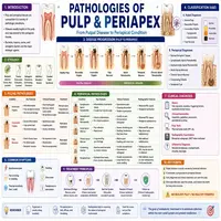
Pulp pathology generally progresses through recognizable stages, each with distinct clinical features. Understanding these stages helps explain why early treatment often saves a tooth that later stages cannot.
- Reversible pulpitis: Mild inflammation causes brief sensitivity to cold, but the tissue can heal once the irritant is removed.
- Irreversible pulpitis: Inflammation becomes severe and self-sustaining, producing spontaneous or lingering pain that does not resolve on its own.
- Pulp necrosis: The tissue dies due to loss of blood supply, often eliminating pain temporarily since nerve function stops.
- Periapical extension: Bacteria and toxins exit through the root apex, initiating inflammation in the surrounding bone.
At each stage, the disease becomes progressively harder to reverse without intervention. Therefore, distinguishing reversible from irreversible pulpitis during examination remains one of the most important diagnostic tasks in endodontics.
From Pulp Disease to Periapical Pathology: A Flowchart
The transition from pulp inflammation to periapical disease follows a fairly predictable sequence. The flowchart below illustrates this progression.
Bacterial Invasion of the Pulp (via caries, trauma, or crack)
        │
        ▼
Reversible Pulpitis (mild, temporary inflammation)
        │
        ▼
Irreversible Pulpitis (persistent, worsening inflammation)
        │
        ▼
Pulp Necrosis (tissue death, loss of blood supply)
        │
        ▼
Bacterial Toxins Exit Through the Root Apex
        │
        ▼
Periapical Inflammation Begins (acute or chronic)
        │
        ▼
Periapical Abscess, Granuloma, or Cyst Forms
This sequence highlights why untreated tooth pain often changes character over time. Initial sharp sensitivity may fade as the pulp dies, only to return later as a dull, persistent ache once periapical tissue becomes involved. Recognizing this pattern helps patients understand why “the pain went away” does not always mean the problem resolved.
Periapical Lesions and Their Characteristics
Once infection reaches the periapex, several distinct lesion types can develop, each with different clinical behavior. The table below compares the most common forms.
| Lesion Type | Nature | Key Feature |
|---|---|---|
| Acute apical periodontitis | Sudden inflammation | Tenderness to biting, minimal radiographic change |
| Chronic apical periodontitis | Long-standing, low-grade inflammation | Often asymptomatic, visible bone changes on X-ray |
| Periapical abscess | Localized collection of pus | Swelling, throbbing pain, possible fever |
| Periapical granuloma | Chronic inflammatory tissue mass | Slow-growing, usually painless |
| Radicular cyst | Fluid-filled sac lined with epithelium | May enlarge gradually, requires surgical or endodontic care |
Radiographic imaging plays a central role in distinguishing these lesions, since granulomas and cysts often appear similar on standard X-rays. In such cases, further evaluation, sometimes including biopsy, helps guide accurate diagnosis and appropriate treatment planning.
Concept Map: The Pathology of Pulp and Periapex
To visualize how these processes connect, consider the concept map below.
                PATHOLOGY OF PULP AND PERIAPEX
                            │
      ┌─────────────────────┼─────────────────────┐
      │                     │                       │
   CAUSES                STAGES                 OUTCOMES
      │                     │                       │
    Caries          Reversible pulpitis        Abscess
    Trauma         Irreversible pulpitis      Granuloma
 Iatrogenic injury      Pulp necrosis        Radicular cyst
Chemical irritation  Periapical extension   Chronic periodontitis
      │                     │                       │
      └─────────────────────┼─────────────────────┘
                            │
                    CLINICAL DIAGNOSIS
                  AND TREATMENT PLANNING
This map demonstrates why endodontic diagnosis relies on connecting cause, disease stage, and resulting lesion type. Missing any link in this chain can lead to incomplete treatment or a misdiagnosed condition.
Diagnosis and Treatment Approaches
Accurate diagnosis depends on combining several sources of information rather than relying on a single test. Clinicians typically use thermal testing, electric pulp testing, percussion testing, and radiographic imaging together to determine whether pulp tissue is still vital and whether periapical involvement has begun.
Treatment decisions follow directly from this diagnosis. Reversible pulpitis, for example, may only require removing the irritant and monitoring healing. Irreversible pulpitis or necrosis, on the other hand, generally requires root canal therapy to remove infected tissue and prevent further spread into the periapex.
In cases involving larger periapical lesions, additional procedures such as apicoectomy or surgical intervention may become necessary. Consequently, early diagnosis remains the single most effective way to avoid these more invasive treatments, since smaller lesions typically respond well to conventional root canal therapy alone.
Conclusion
The pathology of pulp and periapex represents a continuous disease process rather than two unrelated conditions. It begins with minor pulp inflammation and, without timely treatment, can progress through necrosis and into destructive periapical lesions. Recognizing each stage allows clinicians to intervene early, often preserving the natural tooth rather than resorting to extraction.
By understanding how caries, trauma, and other irritants set this process in motion, patients can appreciate why prompt treatment of tooth pain matters so much. Ultimately, protecting pulp health early prevents the more complex and costly periapical disease that develops when infection is left unchecked.
Frequently Asked Questions
Bacterial invasion from untreated tooth decay is the most common trigger, though trauma, cracked teeth, and irritation from dental procedures can also initiate the process.
Yes, if the irritant is removed early, such as through a filling that addresses shallow decay, the pulp can often heal and inflammation subsides.
Pain often fades once the pulp dies, since nerve tissue stops functioning. However, infection frequently continues spreading toward the periapex despite the absence of symptoms.
Standard X-rays often cannot distinguish the two reliably, since both appear as similar radiolucent areas. Biopsy or histological examination may be needed for a definitive diagnosis.
Most periapical lesions caused by pulp necrosis require root canal therapy to remove infected tissue. Larger or persistent lesions may additionally need surgical treatment, such as an apicoectomy.