Pulp and Periradicular Tissues: Anatomy, Disease, and Healing
Every restorative dentist eventually encounters cases where the health of the inner tooth structure determines the entire treatment plan. However, many students and even early-career clinicians struggle to fully grasp how these deep tissues respond to injury and infection. Consequently, understanding pulp and periradicular tissues in detail becomes essential before performing any operative procedure. In this article, we will examine the anatomy of these structures, explain common disease processes, and outline how clinicians diagnose and manage related conditions. Additionally, we will use simple tables, a flowchart, and a concept map to make the information easier to absorb. Therefore, whether you are a dental student or a practicing clinician, this guide should sharpen your understanding of a topic central to everyday practice.
What Are These Structures?
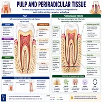
The dental pulp is the soft connective tissue that sits at the center of a tooth, containing blood vessels, nerves, and specialized cells called odontoblasts. Meanwhile, the periradicular region refers to the tissues surrounding the root tip, including the periodontal ligament, cementum, and alveolar bone. Together, these two zones form a functional unit that responds collectively to trauma, decay, and bacterial invasion. As a result, damage in one area frequently triggers a chain reaction affecting the other. Moreover, this close relationship explains why dentists always evaluate both regions together during diagnosis rather than treating them as separate entities.
Furthermore, the pulp plays several biological roles beyond simple sensation. It nourishes the surrounding dentin, produces reparative dentin after injury, and provides an immune response against invading bacteria. Nevertheless, once this tissue becomes severely inflamed or necrotic, its healing capacity drops sharply, which is why early intervention matters so much in clinical practice.
Anatomy Overview
Understanding the structural layout helps clinicians interpret radiographs and plan treatment accurately. The table below summarizes the major components of this anatomical region.
| Structure | Location | Primary Function |
|---|---|---|
| Pulp Chamber | Center of the crown | Houses pulp tissue and nerve endings |
| Root Canal | Extends through the root | Pathway for vessels and nerves |
| Periodontal Ligament | Between root and bone | Anchors tooth, absorbs occlusal forces |
| Cementum | Covers the root surface | Attachment site for ligament fibers |
| Alveolar Bone | Surrounds the root | Provides structural support |
Generally, each of these components works together to maintain tooth vitality and stability. On the other hand, disruption to any single structure can compromise the health of the entire complex over time.
Blood supply also plays a critical role in this anatomical relationship. Small vessels enter through the apical foramen at the root tip, nourishing the pulp and supporting its healing response after injury. Similarly, nerve fibers travel through this same opening, which explains why pulpal pain often feels diffuse and difficult to localize to a single tooth. When trauma or infection disrupts this vascular pathway, the pulp loses its primary source of nutrients and oxygen, accelerating tissue breakdown. Therefore, preserving blood flow through timely treatment often determines whether a tooth remains vital or eventually requires more extensive intervention.
How Disease Progresses: A Flowchart

Bacterial invasion typically follows a predictable path from the outer tooth surface toward the deeper tissues. The flowchart below illustrates this progression in a simplified, step-by-step format.
Flowchart: Pathway of Infection
Step 1: Enamel decay begins at the tooth surface
        |
        v
Step 2: Bacteria penetrate into dentin
        |
        v
Step 3: Toxins reach the pulp chamber
        |
        v
Step 4: Pulp becomes inflamed (pulpitis)
        |
        v
Step 5: Untreated inflammation leads to pulp necrosis
        |
        v
Step 6: Infection spreads to periradicular tissues
        |
        v
Step 7: Periapical lesion or abscess develops
This sequence explains why early detection remains so valuable. Furthermore, catching the problem at the pulpitis stage often lets clinicians save a tooth through conservative treatment rather than extraction. Eventually, if the disease reaches the periradicular stage, more invasive procedures, such as root canal therapy or apical surgery, become necessary.
Common Diseases Affecting Pulp and Periradicular Tissues
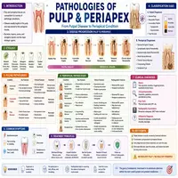
Several distinct conditions can affect this region, each requiring a different diagnostic and treatment approach. Reversible pulpitis, for example, causes mild and short-lived sensitivity that resolves once the irritant is removed. In contrast, irreversible pulpitis produces lingering, spontaneous pain that signals permanent damage to the nerve tissue. Similarly, pulp necrosis occurs when the tissue dies completely, often following untreated irreversible inflammation. Meanwhile, periapical periodontitis develops when infection spreads beyond the root tip into the surrounding bone. In addition, chronic cases may progress into a periapical abscess or, less commonly, a granuloma or cyst. Therefore, accurate diagnosis at each stage directly influences whether the clinician can preserve a tooth through conservative care.
Concept Map: Disease Classification

To visualize how these conditions relate to one another, the table below organizes them under a shared structural theme.
| Level | Category | Sub-Category / Example |
|---|---|---|
| 1 | Dental Pulp-Root Complex | Umbrella term |
| 2 | Pulpal Disease | Reversible Pulpitis, Irreversible Pulpitis, Necrosis |
| 2 | Periradicular Disease | Periapical Periodontitis, Abscess, Granuloma |
| 3 | Clinical Response | Vitality Testing, Radiography, Root Canal Therapy |
This layered structure highlights how pulpal injury, if left unmanaged, naturally progresses toward periradicular involvement. Consequently, clinicians rarely treat these two zones as unrelated problems.
Diagnostic Methods
Accurate diagnosis relies on a combination of clinical tests and imaging. Dentists commonly use thermal testing, electric pulp testing, and percussion testing to assess nerve vitality and inflammation level. Additionally, radiographs reveal periapical changes, bone loss, or widening of the periodontal ligament space that might not be detectable through clinical examination alone. Sometimes, cone-beam computed tomography offers a more detailed three-dimensional view when standard radiographs provide insufficient information. As a result, combining multiple diagnostic tools significantly improves treatment accuracy and reduces the risk of misdiagnosis.
Clinicians also pay close attention to patient history and symptom pattern during evaluation. For instance, sharp pain triggered by cold that lingers after the stimulus disappears often signals irreversible pulpitis rather than a reversible condition. Similarly, spontaneous throbbing pain that worsens at night frequently points toward an already necrotic pulp. Meanwhile, tenderness to biting or percussion suggests that inflammation has already reached the periradicular tissues surrounding the root. Consequently, gathering a detailed symptom history alongside objective test results gives clinicians a much clearer diagnostic picture before any treatment begins.
Treatment Approaches
Treatment selection depends heavily on how far the disease has progressed. For reversible pulpitis, removing the irritant and placing a protective restoration often resolves symptoms completely. However, irreversible pulpitis and necrosis typically require root canal therapy to remove infected tissue and seal the canal system. Afterward, periradicular lesions may need additional intervention, such as apicoectomy, if nonsurgical treatment fails to achieve healing. Importantly, timely treatment preserves natural tooth structure and prevents the need for extraction in most cases. Nonetheless, severely compromised teeth with extensive bone loss may still require extraction despite best efforts.
Prevention and Long-Term Care
Preventing damage to these tissues remains far easier than treating advanced disease. Regular dental checkups allow clinicians to catch decay before it reaches the pulp, which significantly reduces the likelihood of needing invasive treatment later. In addition, prompt attention to cracked teeth, trauma, or deep restorations helps protect the pulp from unnecessary irritation. Patients who maintain good oral hygiene and avoid prolonged exposure to sugary or acidic foods also lower their overall risk of pulpal disease. Afterward, once treatment is complete, periodic follow-up radiographs help confirm that periradicular healing is progressing as expected. Ultimately, a proactive approach benefits both the patient and the long-term success of any restorative work performed.
Conclusion
The health of pulp and periradicular tissues lies at the heart of successful operative and endodontic dentistry. From their intertwined anatomy to the predictable progression of disease, understanding this relationship helps clinicians make timely, accurate decisions. Although early-stage conditions often respond well to conservative care, advanced disease demands more comprehensive intervention to save the tooth. Ultimately, mastering the diagnosis and management of these tissues equips dental professionals to protect long-term oral health and preserve natural dentition whenever possible.
Frequently Asked Questions
The pulp sits inside the tooth and contains nerves and blood vessels, while the periradicular region surrounds the root tip and supports the tooth structurally.
Reversible pulpitis can heal once the irritant is removed, but irreversible pulpitis cannot recover without professional intervention such as root canal therapy.
Clinicians typically combine radiographs, percussion testing, and vitality tests to determine whether infection has spread beyond the pulp into surrounding bone.
Not always; mild, reversible cases may only need a protective restoration, while irreversible damage generally requires root canal therapy.
Untreated infection can progress into a periapical abscess or cyst, potentially leading to bone loss and eventual tooth loss if not managed properly.