The Structure of Skeletal Muscles Explained Simply
Have you ever wondered how your body lifts, walks, or runs? Skeletal muscles make all of this possible. These muscles attach to bones and pull them into motion whenever you move.
Understanding the structure of skeletal muscles helps you appreciate how movement actually happens. In fact, this knowledge benefits athletes, students, and curious readers alike. Additionally, it explains why exercise strengthens the body over time. So, let’s break down this fascinating tissue, layer by layer.
Muscle tissue may look simple from the outside. However, several organized layers work together beneath the surface. Therefore, learning each layer step by step makes the whole picture much clearer.
Movement depends on far more than just muscle size. In fact, the internal arrangement of fibers and filaments determines how efficiently force transfers to bone. Consequently, understanding this arrangement explains why some muscles handle heavy loads while others manage fine, precise motions.
Interestingly, muscle tissue also communicates constantly with the nervous system. Signals travel rapidly between nerves and fibers, coordinating even the smallest movements. As a result, actions like blinking or lifting a cup feel effortless, despite the complex activity happening underneath.
Basic Organization of Muscle Tissue
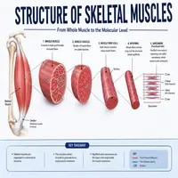
Skeletal muscle starts as a whole organ wrapped in a tough layer called the epimysium. This outer covering protects the entire muscle. Meanwhile, it also separates one muscle from neighboring tissue.
Inside the epimysium, bundles of fibers group together. Each bundle is called a fascicle. A layer named the perimysium surrounds every fascicle. Consequently, this layer allows nerves and blood vessels to reach deep inside the muscle.
Within each fascicle, individual muscle fibers sit side by side. A thin layer called the endomysium wraps each fiber. This delicate covering supports capillaries that feed oxygen and nutrients directly to the cell.
Every muscle fiber is actually a long, cylindrical cell. Numerous nuclei sit along its length, since the fiber forms from multiple cells fusing together. Inside this cell, thousands of smaller strands called myofibrils run in parallel.
Notably, muscle fibers vary in type as well. Slow-twitch fibers resist fatigue and support endurance activities. Fast-twitch fibers, on the other hand, generate quick bursts of power but tire more rapidly. Most muscles contain a mixture of both types, depending on their overall function.
This nested design is central to the structure of skeletal muscles. Each layer, from the whole muscle down to individual fibers, adds strength and organization. As a result, the tissue can generate powerful, coordinated movement.
Inside the Muscle Fiber: Myofibrils and Filaments
Myofibrils fill most of the space inside each muscle fiber. These thread-like structures contain repeating units called sarcomeres. Sarcomeres are the basic functional units responsible for contraction.
Two types of protein filaments make up each sarcomere. Thick filaments consist mainly of myosin. Thin filaments, on the other hand, consist mainly of actin. These filaments overlap in a precise pattern, creating the characteristic striped appearance under a microscope.
Additionally, other proteins support this system. Titin acts like a spring, keeping filaments aligned. Troponin and tropomyosin regulate when filaments can interact. Together, these proteins ensure smooth, controlled contraction.
Furthermore, the sarcoplasmic reticulum surrounds each myofibril like a mesh. This structure stores calcium ions until a nerve signal triggers their release. Similarly, tiny tunnels called T-tubules carry electrical signals deep into the fiber, ensuring every part contracts at the same moment.
How Skeletal Muscles Contract
Contraction involves a clear sequence of events. Here is a simple flowchart of the process:
Nerve signal arrives
      ↓
Calcium releases into the cell
      ↓
Calcium binds to troponin
      ↓
Myosin heads attach to actin
      ↓
Filaments slide past each other
      ↓
Sarcomere shortens
      ↓
Muscle contracts
First, a nerve impulse reaches the muscle fiber. Then, calcium ions flood out of internal storage. Meanwhile, these ions bind to troponin, shifting tropomyosin out of the way.
Next, myosin heads latch onto exposed sites on actin filaments. Subsequently, they pull the thin filaments inward using energy from ATP. This sliding motion shortens each sarcomere.
Finally, thousands of sarcomeres shorten simultaneously along the fiber. As a result, the entire muscle contracts and generates force. Once the nerve signal stops, calcium returns to storage, and the muscle relaxes.
This entire cycle repeats rapidly with every movement you make. Whether you are climbing stairs or typing a message, countless sarcomeres fire in coordinated waves. Truly, the speed and precision of this process highlight just how remarkable muscle physiology really is.
Key Components and Their Functions
For clarity, here is a quick reference table of the main structural components.
| Component | Location | Main Function |
|---|---|---|
| Epimysium | Outer covering | Protects the whole muscle |
| Perimysium | Around fascicles | Bundles fibers and carries vessels |
| Endomysium | Around each fiber | Supports capillaries and nerves |
| Myofibril | Inside fiber | Contains contractile units |
| Sarcomere | Inside myofibril | Basic unit of contraction |
| Myosin | Thick filament | Pulls actin during contraction |
| Actin | Thin filament | Slides past myosin to shorten fiber |
This table highlights how each layer supports the next. Indeed, damage to any single layer can affect overall muscle performance.
Beyond these core parts, blood vessels and nerve endings weave through every layer. Oxygen and nutrients travel through capillaries reaching each fiber. Meanwhile, nerve endings deliver the signals that initiate every contraction, linking structure directly to function.
Common Conditions Affecting Muscle Tissue
Unfortunately, several conditions can disrupt normal muscle function. Muscular dystrophy, for example, weakens fibers due to genetic mutations affecting structural proteins. Over time, this condition reduces strength and mobility.
Myopathy refers to a broader group of disorders affecting muscle tissue directly. Symptoms often include weakness, cramping, or fatigue. Meanwhile, rhabdomyolysis occurs when muscle fibers break down rapidly, releasing harmful proteins into the bloodstream.
Muscle strains are also extremely common, especially among athletes. This injury happens when fibers stretch or tear beyond their normal limit. Similarly, atrophy develops when muscles shrink from disuse, illness, or prolonged immobility.
Tendinitis is another frequent concern, though it technically affects the connective tissue near muscles. Repetitive motion often triggers inflammation and discomfort. Meanwhile, cramps occur when a muscle contracts involuntarily and fails to relax right away.
Fortunately, most muscle issues improve with proper treatment. Physical therapy, rest, and gradual strengthening exercises often restore function effectively.
Tips to Keep Skeletal Muscles Healthy
Good habits protect this tissue throughout life. Below are practical steps anyone can follow.
- Exercise regularly with both strength and cardio training.
- Eat sufficient protein to support fiber repair and growth.
- Stay hydrated, since water supports every cellular process.
- Stretch before and after workouts to reduce injury risk.
- Get adequate sleep, because tissue repair happens during rest.
- Avoid sudden, excessive strain on untrained muscles.
Moreover, gradual progression during training prevents unnecessary tears. Likewise, warming up increases blood flow and prepares fibers for activity. Furthermore, balanced nutrition supplies the building blocks fibers need to rebuild stronger after exercise.
Additionally, rest days matter just as much as workout days. Muscles repair and grow during recovery, not during the activity itself. Therefore, skipping rest can slow progress and increase injury risk over time.
Conclusion
The structure of skeletal muscles reveals an incredibly organized system. From the outer epimysium down to individual actin and myosin filaments, every layer plays a specific role. As we have seen, this organization allows for powerful, precise movement throughout the body.
Understanding this structure helps explain why proper nutrition, exercise, and rest matter so much. Furthermore, recognizing early signs of muscle problems can prevent long-term complications. So, take a moment to appreciate the remarkable design behind every step, lift, and movement you make each day.
Frequently Asked Questions
The sarcomere is the basic unit of contraction. It contains overlapping actin and myosin filaments that slide past each other to shorten the fiber.
A thin layer called the endomysium covers each muscle fiber. This layer supports capillaries that deliver oxygen and nutrients directly to the cell.
Calcium binds to troponin, which shifts tropomyosin away from binding sites. This action allows myosin heads to attach to actin and begin contraction.
Muscle strains occur when fibers stretch or tear beyond their normal capacity. Common causes include sudden movements, overexertion, or inadequate warm-up.
Regular strength training, adequate protein intake, and consistent activity help maintain muscle mass with age. Additionally, staying active reduces the risk of atrophy and weakness.