The Eye: A Complete Guide to Its Anatomy and Function
Have you ever wondered how you see the world around you? The eye makes it possible. This small, round organ captures light, turns it into signals, and sends those signals to your brain. Consequently, you experience color, depth, and movement in an instant.
Understanding this organ is not just for doctors or students. In fact, everyone benefits from knowing how their vision works. Additionally, this knowledge helps you spot problems early and protect your sight for years to come. So, let’s explore the anatomy of this incredible structure, piece by piece.
Vision shapes almost every part of daily life. From reading a book to recognizing a friend across the street, this sense guides countless decisions each day. Therefore, learning how it functions is more than an academic exercise. It is a practical step toward better health.
Interestingly, this organ works closely with the brain, not alone. Signals travel back and forth constantly, and the brain fills in gaps whenever needed. As a result, vision feels seamless, even though the underlying process is complex.
The Basic Structure of This Organ
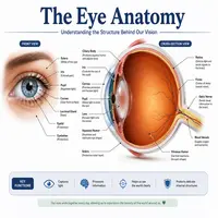
The eye sits inside a bony socket called the orbit. This socket protects it from injury. Meanwhile, six small muscles attach to the outer surface and allow it to move in every direction.
The outer layer consists of the sclera and cornea. The sclera is the white, tough coating that gives the eyeball its shape. The cornea, on the other hand, is the clear, dome-shaped front section. It bends light as it enters.
Behind the cornea lies the iris, the colored ring around the pupil. The iris controls the size of the pupil, the black opening in its center. Therefore, it regulates how much light reaches the back of the eyeball.
Next comes the lens. This flexible structure sits just behind the iris. It fine-tunes focus by changing shape. As a result, you can see objects clearly whether they are near or far.
Finally, the retina lines the back wall. This thin layer contains millions of light-sensitive cells. These cells, called rods and cones, convert light into electrical signals. Afterward, the optic nerve carries those signals to the brain.
Between these structures, two fluids maintain shape and clarity. Aqueous humor fills the front chamber, while vitreous humor fills the larger back chamber. Together, they keep pressure balanced and nourish nearby tissues.
Additionally, the choroid layer sits between the retina and sclera. This layer supplies blood and oxygen to surrounding tissue. Without it, the retina could not function properly for long.
How Vision Works: Step by Step
Vision seems instant, yet several steps happen every time you look at something. Here is a simple flowchart of the process:
Light rays enter
      ↓
Cornea bends the light
      ↓
Pupil controls light amount
      ↓
Lens fine-tunes the focus
      ↓
Retina captures the image
      ↓
Optic nerve sends signals
      ↓
Brain interprets the image
First, light bounces off an object and travels toward you. Then, the cornea bends this light so it can travel deeper. Meanwhile, the iris adjusts the pupil size based on brightness. In bright rooms, the pupil shrinks. In dim rooms, it widens.
Next, the lens focuses the light precisely onto the retina. This step is called accommodation. Subsequently, rod and cone cells in the retina transform the light into nerve impulses. Rods handle low light and movement, while cones handle color and fine detail.
Finally, the optic nerve transmits these impulses to the visual cortex in the brain. Within milliseconds, your brain assembles the signals into a clear picture. Truly, this entire chain works so smoothly that you rarely notice it happening.
Notably, having two working eyes also creates depth perception. Each side captures a slightly different angle of the same scene. Subsequently, the brain merges both images, producing a three-dimensional view of the world. This is why closing one eye can make judging distance more difficult.
Key Parts and Their Functions
For clarity, here is a quick reference table of the main components and their roles.
| Part | Location | Main Function |
|---|---|---|
| Cornea | Front surface | Bends incoming light |
| Iris | Behind cornea | Controls pupil size and light entry |
| Pupil | Center of iris | Opening that lets light pass through |
| Lens | Behind iris | Focuses light onto the retina |
| Retina | Back wall | Converts light into nerve signals |
| Optic Nerve | Rear of eyeball | Sends signals to the brain |
| Sclera | Outer coating | Protects and shapes the eyeball |
| Vitreous Humor | Middle chamber | Maintains shape and clarity |
This table shows how each part depends on the others. Indeed, if one piece fails, the entire system struggles to produce clear vision.
Common Conditions That Affect Vision
Unfortunately, several conditions can disrupt normal function. Nearsightedness, for example, occurs when light focuses in front of the retina rather than on it. Farsightedness happens the opposite way.
Astigmatism results from an irregularly shaped cornea. Consequently, images appear blurry or stretched at every distance. Meanwhile, cataracts develop when the lens becomes cloudy over time, often with age.
Glaucoma is another common concern. This condition raises pressure inside the eyeball, and it can damage the optic nerve. Similarly, macular degeneration affects the retina and reduces central vision, particularly in older adults.
Dry eye syndrome is also widespread, especially among screen users. This condition occurs when tear production drops or tears evaporate too quickly. Consequently, symptoms include irritation, redness, and blurred vision.
Floaters, meanwhile, appear as small specks drifting across your view. Usually, they result from natural changes in the vitreous humor with age. However, a sudden increase in floaters should prompt an immediate checkup.
Fortunately, most of these conditions respond well to early treatment. Regular checkups, therefore, play a huge role in preserving long-term vision.
Simple Tips to Protect Your Vision
Good habits go a long way toward keeping this organ healthy. Below are practical steps anyone can follow.
- Schedule an eye exam every one to two years.
- Wear sunglasses that block UV rays outdoors.
- Follow the 20-20-20 rule during screen time: every 20 minutes, look at something 20 feet away for 20 seconds.
- Eat foods rich in vitamin A, C, and E, along with omega-3 fatty acids.
- Avoid rubbing your eyes, since this can irritate delicate tissue.
- Stay hydrated, because dryness often leads to discomfort and blurred vision.
Moreover, quitting smoking significantly lowers the risk of cataracts and macular degeneration. Likewise, managing conditions like diabetes and high blood pressure protects the retina’s blood vessels.
Furthermore, proper lighting reduces strain while reading or working. Dim rooms often force the lens to work harder, which leads to fatigue over time. Similarly, getting enough sleep allows tissues to repair and refresh naturally each night.
Finally, protective eyewear matters during sports or manual labor. Sudden impacts can cause serious damage in an instant. Therefore, a small precaution now can prevent a lifetime of complications later.
Conclusion
The eye is truly one of the body’s most fascinating organs. From the cornea to the optic nerve, every part works together in perfect harmony. As we have seen, light travels through several structures before your brain finally interprets an image.
Understanding this process helps you appreciate how precious your vision really is. Furthermore, small daily habits, such as wearing sunglasses or scheduling regular exams, can prevent many common problems. So, take a moment today to care for this organ that lets you see the world so clearly.
Frequently Asked Questions
Its main function is to capture light and convert it into signals the brain can interpret as images. This process allows you to see shapes, colors, and movement.
The cornea and lens work together to focus light. The cornea does most of the bending, while the lens fine-tunes the focus for near and far objects.
The pupil changes size to control how much light enters. It shrinks in bright conditions and widens in dim conditions, thanks to the iris muscles.
Blurry vision often results from refractive errors like nearsightedness, farsightedness, or astigmatism. Cataracts and other conditions can also cause blurriness, especially with age.
Most experts recommend an exam every one to two years for healthy adults. However, people with existing conditions or a family history of eye disease may need more frequent checkups.