Prevertebral Region: A Complete Anatomy Guide
The neck holds many hidden structures. One of the most important, yet often overlooked, is the prevertebral region. This area sits deep within the neck, right in front of the cervical spine. It plays a key role in posture, neck movement, and protecting nearby nerves and vessels.
In this guide, we will break down the anatomy of this region in simple terms. We will also look at its muscles, fascia, and clinical relevance. Whether you are a student or a healthcare professional, this article will help you understand the topic clearly.
What Is the Prevertebral Region?
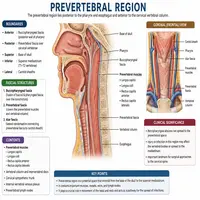
The prevertebral region refers to the space located anterior to the vertebral bodies of the cervical spine. It extends from the base of the skull down to the upper thoracic area. This region houses several deep muscles, along with a fascial covering that separates it from other neck compartments.
Because of its central position, this area connects many important structures. Nerves, blood vessels, and muscles all pass through or near it. Therefore, understanding its layout helps explain many neck-related conditions.
Additionally, the region acts as a support system for the cervical spine. It stabilizes the neck during movements like flexion and rotation. Without it, everyday actions such as turning your head would feel unstable.
Location and Boundaries
This area lies deep to the prevertebral fascia, which wraps around the muscles like a sleeve. Above, it connects to the base of the skull. Below, it continues into the thorax, blending with structures near the diaphragm.
On each side, the region borders the carotid sheath. This sheath contains the carotid artery, jugular vein, and vagus nerve. Consequently, any swelling or infection in this space can affect these vital structures.
The boundaries can be summarized as follows:
| Boundary | Structure |
|---|---|
| Superior | Base of the skull |
| Inferior | Thoracic cavity (near T3 level) |
| Anterior | Prevertebral fascia |
| Posterior | Vertebral bodies and longus muscles |
| Lateral | Carotid sheath on both sides |
This table gives a quick snapshot of how the space is framed within the neck.
Prevertebral Fascia: Structure and Layers
The prevertebral fascia is a thin but tough layer of connective tissue. It covers the deep muscles of the neck and spine. Think of it as a protective wrapper that keeps these muscles in place.
This fascia has two main parts. The alar fascia lies more anteriorly, while the true prevertebral layer sits closer to the vertebral column. Between these layers, a potential space exists called the “danger space.” This space is clinically significant because infections can spread through it quickly, reaching down into the chest cavity.
Moreover, the fascia attaches firmly to the transverse processes of the cervical vertebrae. This attachment gives it strength and stability. As a result, it helps contain infections and limits their spread in many cases, though not always completely.
Muscles Within the Prevertebral Compartment
Several muscles occupy this deep neck compartment. These muscles work together to flex the neck and support the head.
Here is a simple breakdown:
| Muscle | Main Action | Nerve Supply |
|---|---|---|
| Longus colli | Flexes and rotates the neck | Cervical spinal nerves (C2–C6) |
| Longus capitis | Flexes the head | Cervical spinal nerves (C1–C3) |
| Rectus capitis anterior | Flexes the head at atlanto-occipital joint | C1 spinal nerve |
| Rectus capitis lateralis | Stabilizes the head laterally | C1 spinal nerve |
These muscles rarely get much attention, yet they perform essential daily functions. For instance, nodding your head or tilting it sideways relies heavily on this muscle group.
Contents and Neurovascular Structures
Beyond muscles, this space also contains important nerve branches and small vessels. The cervical sympathetic trunk runs along the surface of these muscles. This trunk plays a role in regulating blood vessel size and sweat gland activity in the head and neck.
Additionally, small branches from the cervical plexus travel through this area. These branches supply sensation and motor control to nearby structures. Meanwhile, the ascending cervical artery, a branch of the inferior thyroid artery, provides blood supply to these muscles.
Because so many structures pass through this compact space, even small changes in anatomy can cause noticeable symptoms. For example, compression here might lead to neck stiffness or nerve-related discomfort.
Prevertebral Space and Its Clinical Significance
Understanding the prevertebral region matters greatly in clinical practice. Infections, abscesses, and trauma can all affect this space. Since it connects to the mediastinum, infections here can travel downward and cause serious complications.
Below is a simple flowchart showing how an infection might spread through this region:
Neck Infection (e.g., dental or pharyngeal origin)
        ↓
Spreads into Prevertebral Space
        ↓
Travels along Danger Space
        ↓
Reaches Posterior Mediastinum
        ↓
Possible Mediastinitis (Emergency)
This flowchart highlights why early diagnosis matters so much. A minor throat infection, if left untreated, could potentially become life-threatening.
Furthermore, trauma to the cervical spine can cause bleeding into this space. This bleeding may push the pharynx forward, leading to difficulty swallowing or breathing. Therefore, doctors often check this area carefully during neck trauma evaluations.
Common Disorders Affecting This Area
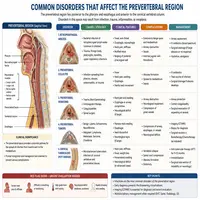
Several conditions are linked to this deep neck compartment. Below are some of the most common ones:
- Retropharyngeal abscess – often spreads into the prevertebral space in severe cases.
- Prevertebral hematoma – usually caused by trauma or fractures.
- Tuberculous spondylitis – can lead to abscess formation in this region.
- Cervical spine infections – may extend into surrounding soft tissue.
Each of these conditions requires prompt medical attention. Delayed treatment can lead to airway compromise or spread of infection.
Diagnostic Imaging Approaches
Doctors typically use imaging to assess this deep neck space. CT scans with contrast remain the gold standard for detecting fluid collections or abscesses. MRI, on the other hand, offers better soft tissue detail, especially when nerve involvement is suspected.
X-rays can sometimes show widening of the space between the trachea and spine, hinting at swelling. However, they are less detailed compared to CT or MRI.
In emergency settings, quick imaging helps guide treatment decisions. Since infections here can progress rapidly, timely diagnosis often saves lives.
Conclusion
The prevertebral region may be small, but its role is significant. It houses key muscles, protects vital nerves, and connects the neck to the chest. Understanding its anatomy helps explain many clinical conditions, from infections to trauma-related injuries.
For students, mastering this topic builds a strong foundation in head and neck anatomy. For clinicians, recognizing its boundaries and contents supports faster, safer diagnosis. Overall, this compact space plays a much bigger role than its size suggests.
Frequently Asked Questions
It is the deep space in the neck located just in front of the cervical spine. It contains muscles, nerves, and a protective fascial layer.
This fascia wraps around deep neck muscles and helps contain infections, although it cannot always stop their spread completely.
Yes. Through the danger space, infections can travel down into the posterior mediastinum, leading to serious complications.
The main muscles include longus colli, longus capitis, rectus capitis anterior, and rectus capitis lateralis.
CT scans with contrast are commonly used, while MRI provides better detail for soft tissue and nerve involvement.