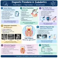
Diagnostic Procedures in Endodontics Explained
Tooth pain rarely tells the whole story. A dull ache might come from the pulp, the gum, or even a sinus. That is why careful diagnosis matters. In this guide, we walk through the diagnostic procedures in endodontics step by step. You will learn what your dentist asks, checks, and measures. Along the way, you will find a flowchart and two handy tables. Think of your dentist as a detective. Each test adds one more clue. Together, those clues point to the right treatment.
Why Diagnostic Procedures in Endodontics Matter
Accurate diagnosis protects your tooth. Without it, treatment becomes guesswork. For example, a cracked tooth and a gum infection can feel identical. However, each one needs a very different fix. Treating the wrong problem wastes time and money. Worse, it leaves the real cause untouched. Every clue counts. Symptoms, tests, and images each play a part. Alone, none of them settles the matter.
Good testing also spares healthy teeth. Sometimes pain seems to come from an upper tooth. Yet it starts in a lower one. Dentists call this referred pain. It confuses patients and dentists alike. As a result, your dentist never relies on one test alone. Instead, they combine several findings. Then they compare those findings with your symptoms. Consequently, the final answer rests on solid evidence.
Timing matters too. Early diagnosis often saves the tooth. Delays let infection spread into the bone. Meanwhile, a quick and accurate workup keeps treatment simple. Most importantly, you stay informed at every stage. Let’s start where every diagnosis begins, with your story.
Step One: Listening to Your Story
Every diagnosis starts with a conversation. Your dentist wants to hear about your pain in your own words. First, they ask when it began. Then they ask what triggers it. Does hot coffee make it worse? Maybe cold water brings relief instead. Each answer narrows the possibilities.
Pain quality matters too. Sharp, brief pain often points to inflamed pulp that can still heal. In contrast, lingering throbbing usually signals deeper damage. Pain that wakes you at night deserves special attention. Likewise, pain when you bite suggests trouble around the root.
Your dentist also reviews your medical history. Conditions like diabetes can slow healing. Certain medicines change how your body fights infection. Therefore, be honest about everything you take. Previous dental work counts as well. A deep filling or old crown may hide new decay. Also mention any recent injury to your face or mouth. Even a small knock can damage a nerve months later. With this background, your dentist moves on to the exam.
Clinical Examination: A Close Look Inside Your Mouth
Next comes the visual exam. Your dentist checks your teeth, gums, and soft tissues. They look for swelling, redness, and small pimple-like bumps on the gum. That bump is a sinus tract. It drains infection from deep inside the bone. Therefore, it acts as a strong clue.
Then they study the tooth itself. Cracks, deep cavities, and worn fillings all raise concern. Sometimes a tooth looks darker than its neighbors. That color change often signals a dying nerve. Meanwhile, a tiny crack can hide from the naked eye. To find it, your dentist may use magnification and special dyes.
Gum health gets attention too. Using a thin probe, they measure the space around each tooth. Deep pockets can point to gum disease instead of a nerve problem. In some cases, both problems exist together. Because of that, the probe readings matter. Finally, your dentist gently presses and taps the area. Tenderness here suggests inflammation near the root tip.
Pulp Sensibility Tests: Cold, Heat, and Electric
Next, your dentist tests how your nerve responds. These tests sound scary, but they feel brief. Most patients describe a quick zing, nothing more. The response tells your dentist whether the pulp is alive and healthy.
Cold testing comes first. Your dentist touches a cold cotton pellet to the tooth. A healthy tooth reacts for a second or two, then calms down. However, a tooth that keeps hurting needs a closer look. A tooth with no response may have lost its nerve.
Now the heat test works in reverse. Heat can reveal problems that cold misses. Dentists use it carefully, because heat can harm sensitive tissue. Also, the electric pulp test sends a tiny current into the tooth. You raise a hand when you feel tingling. Higher numbers mean the nerve has less life left.
To keep results reliable, your dentist always tests a healthy tooth first. That gives a baseline for comparison. Below, you will find a summary of the main tests.
| Test | What It Checks | Typical Finding |
|---|---|---|
| Cold test | Nerve vitality | Brief zing means healthy; lingering pain means inflamed pulp |
| Heat test | Deep pulp inflammation | Sharp, lasting pain suggests irreversible damage |
| Electric pulp test | Nerve response | No response suggests a non-vital tooth |
| Percussion (tapping) | Root tip inflammation | Pain on tapping points to apical inflammation |
| Palpation (pressing) | Tissue tenderness | Tender gum suggests infection near the root |
| Bite test | Cracks in the tooth | Pain on release hints at a fracture |
X-Rays and Advanced Imaging
Tests tell one story, and images tell another. X-rays reveal what eyes cannot see. A small periapical film shows the whole tooth, from crown to root tip. On it, your dentist looks for dark shadows near the root. Those shadows often mean bone loss from infection. They also check canal shape, old fillings, and root length.
However, a flat X-ray has limits. It squashes a three-dimensional tooth into two dimensions. Overlapping structures can hide problems. For this reason, dentists sometimes order a cone-beam CT scan. This scan builds a 3D picture of the tooth and bone. As a result, it reveals extra canals, cracks, and tiny lesions.
Still, dentists do not order it for every patient. The scan delivers more radiation than a standard film. So your dentist weighs the benefit carefully. Most routine cases need only regular X-rays. Complicated cases, such as retreatments, gain the most from the 3D view. Once every clue arrives, your dentist connects the dots.
Special Situations: Cracked and Previously Treated Teeth
Some cases need extra detective work. Cracked teeth top the list. A crack can cause pain only when you release a bite. Meanwhile, the tooth may look perfectly normal on an X-ray. To catch it, your dentist uses a small bite stick. You bite down on it, one tooth at a time. Pain on release often points to the culprit.
Additionally, dentists may shine a bright light through the tooth. Cracks block the light and show up as dark lines. Staining dyes can highlight them too. Once found, the crack’s depth decides the outlook. Shallow cracks often respond well to a crown. Deep cracks that reach the root may not.
Teeth with old root canals raise different questions. Sometimes pain returns years later. In that case, your dentist looks for missed canals or new leakage. Old fillings can also hide fresh decay. Therefore, X-rays and a careful history become even more important. A specialist may join the process here. Fortunately, modern microscopes make these hidden problems much easier to spot.
Children need special care as well. Their teeth have wide canals and open root tips. Consequently, test responses can differ from adults. Dentists rely more on swelling, mobility, and X-rays for young patients. They also keep exams gentle and short.
Preparing for Your Diagnostic Visit
A little preparation makes your appointment smoother. First, write down your symptoms. Note when the pain starts, how long it lasts, and what eases it. Next, bring a list of your medicines. Also mention any allergies, especially to anesthetics.
Try not to take painkillers right before your visit. They can mask helpful clues. However, never suffer through severe pain just to prove a point. Call your dentist and explain the situation. Finally, arrive relaxed. Deep breaths help, and so does asking questions. A calm patient makes testing easier and results clearer.
From Symptoms to Diagnosis: A Simple Flowchart
Here is how the pieces fit together. Notice that each step builds on the one before it.
        Patient reports tooth pain
                   |
                   v
      Medical and dental history review
                   |
                   v
    Visual exam, probing, and palpation
                   |
                   v
         Percussion and bite tests
                   |
                   v
     Cold, heat, and electric pulp tests
                   |
                   v
    Periapical X-ray (CBCT if needed)
                   |
                   v
           Combine all findings
                   |
        +----------+-----------+
        |                      |
        v                      v
  Pulp healthy or       Pulp damaged beyond
  able to recover            repair
        |                      |
        v                      v
  Monitor or place       Root canal treatment
  a simple filling
Common Endodontic Diagnoses
Dentists sort their findings into clear labels. Each label guides the next move. Also, these labels give your dental team a shared language. The table below shows the most common ones.
| Diagnosis | Typical Symptoms | Usual Treatment |
|---|---|---|
| Reversible pulpitis | Brief pain to cold or sweets | Filling and monitoring |
| Irreversible pulpitis | Lingering, spontaneous pain | Root canal treatment |
| Pulp necrosis | No response; sometimes a darker tooth | Root canal treatment |
| Acute apical abscess | Swelling, severe pain, pain on biting | Drainage and root canal |
| Chronic apical periodontitis | Little pain; shadow on the X-ray | Root canal or retreatment |
Conclusion
Good endodontic diagnosis takes patience and teamwork. Your dentist listens to your story, examines your mouth, and runs simple tests. Then X-rays add the final pieces. Because no single test tells everything, each clue matters. In the end, this careful process protects you from unnecessary treatment. It also helps save teeth that might otherwise need removal. So, if a tooth hurts, book a visit early. A quick, thorough workup gives you clear answers and a confident plan.
Frequently Asked Questions
Not really. Most patients feel a brief zing or tingle. The sensation fades within seconds. Your dentist can pause anytime you ask.
Tapping checks the tissue around the root. Pain here suggests inflammation or infection. Healthy teeth usually feel nothing at all.
No. Standard X-rays flatten a 3D structure, so small cracks or extra canals can hide. That is why dentists combine X-rays with other tests. In tricky cases, a 3D scan adds detail.
Most exams take 30 to 45 minutes. Emergency visits often run shorter. Complex cases may need a second appointment or a specialist referral.
Not always. Reversible pulpitis often heals with a simple filling. Your dentist recommends a root canal only when the pulp cannot recover.