Nerve Impulse and Neuromuscular Junction: How Signals Move Muscles
Every move you make starts with a spark. A nerve cell fires, and that spark travels down a long fiber until it reaches a muscle. This handoff point is where the impulse and neuromuscular junction do their real work. Without this tiny gap, your muscles would never receive the order to contract. In this article, we will break down the science in plain language. We will also cover the structure involved, the step-by-step process, common disorders, and quick-reference tables you can use for study or teaching.
What Is a Nerve Impulse?
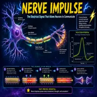
A nerve impulse is an electrical signal. It travels along a neuron from the cell body to the axon terminal. This signal is also called an action potential, and it depends on the movement of charged particles across the neuron’s membrane.
Here is how it happens, step by step:
- A stimulus triggers sodium channels to open.
- Sodium ions rush into the neuron, and the inside becomes more positive.
- This shift, called depolarization, spreads along the axon.
- Potassium channels then open, and potassium exits the cell.
- The membrane returns to its resting state, a process known as repolarization.
Meanwhile, this entire cycle happens in milliseconds. As a result, a single neuron can fire hundreds of times per second. Consequently, muscles can respond almost instantly to a decision made in the brain.
Speed matters here. Myelin, a fatty sheath around many axons, speeds up conduction significantly. Therefore, myelinated neurons carry impulses much faster than unmyelinated ones. This is why damage to myelin, as seen in multiple sclerosis, slows or blocks signals.
Before any of this can happen, though, the neuron must sit at rest. At rest, the inside of the membrane holds a negative charge compared to the outside. This resting potential, usually around -70 millivolts, sets the stage for the next signal. Sodium-potassium pumps maintain this balance by moving three sodium ions out for every two potassium ions they bring in. Without this constant pumping, the neuron would lose its ability to fire altogether.
Once the impulse reaches the end of the axon, it cannot simply jump into the muscle. Instead, it needs a translator. That translator is the neuromuscular junction, and this is exactly where our next section begins.
The Neuromuscular Junction: Structure Explained
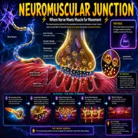
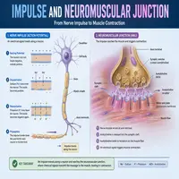
The neuromuscular junction (NMJ) is the meeting point between a motor neuron and a skeletal muscle fiber. It is not a direct physical connection. Instead, a small gap called the synaptic cleft separates the two.
Three main components make up this junction:
| Component | Location | Main Function |
|---|---|---|
| Presynaptic terminal | End of the motor neuron | Stores and releases acetylcholine |
| Synaptic cleft | Space between neuron and muscle | Allows neurotransmitter diffusion |
| Motor end plate | Muscle fiber membrane | Contains receptors for acetylcholine |
Additionally, the presynaptic terminal holds vesicles filled with acetylcholine (ACh). These vesicles wait near the membrane until they receive a signal to release their contents. Meanwhile, the motor end plate is folded into many small ridges. This folding increases surface area, so more receptors can bind ACh at once.
Because this structure is so specialized, even small disruptions can cause major problems. For instance, autoimmune attacks on ACh receptors lead to serious muscle weakness, which we will discuss later in this article.
How the Impulse and Neuromuscular Junction Work Together
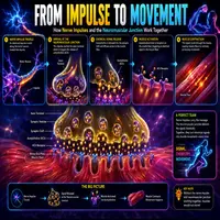
Now let’s connect the two systems. The impulse and neuromuscular junction work as a relay team, passing a signal from nerve to muscle in a precise sequence.
Below is a simplified flowchart of the process:
Nerve impulse reaches axon terminal
            |
            v
Calcium channels open
            |
            v
Calcium enters the presynaptic terminal
            |
            v
Vesicles release acetylcholine into synaptic cleft
            |
            v
Acetylcholine binds to receptors on motor end plate
            |
            v
Sodium channels open on muscle membrane
            |
            v
Muscle action potential begins
            |
            v
Calcium releases inside muscle fiber
            |
            v
Muscle contraction occurs
First, the nerve impulse arrives at the axon terminal. Next, voltage-gated calcium channels open in response. Calcium then flows into the neuron, and this triggers vesicles to fuse with the membrane. Subsequently, acetylcholine spills into the synaptic cleft and diffuses across the gap.
Once acetylcholine reaches the motor end plate, it binds to nicotinic receptors. This binding opens sodium channels on the muscle membrane, and a new action potential begins—this time in the muscle itself. Afterward, this electrical signal spreads across the muscle fiber and triggers calcium release inside the cell. Finally, the muscle contracts.
Enzymes called acetylcholinesterase quickly break down ACh in the cleft. This step is critical. Otherwise, the muscle would stay in a constant state of stimulation, and relaxation would become impossible.
Clinical Relevance: When the Junction Fails
Several conditions target the impulse and neuromuscular junction directly, and understanding them highlights just how important this system is.
Myasthenia gravis is one well-known example. In this disorder, the immune system attacks ACh receptors on the motor end plate. As a result, fewer receptors remain available, and muscle signals weaken over the course of repeated use. Patients often notice drooping eyelids, fatigue, and weakness that worsens throughout the day.
Lambert-Eaton myasthenic syndrome works differently. Here, antibodies attack the calcium channels on the presynaptic terminal instead. Consequently, less acetylcholine gets released, though the effect can sometimes improve slightly with repeated activity.
Botulinum toxin, meanwhile, blocks acetylcholine release entirely. This is why botulism causes severe paralysis, yet in controlled medical doses, it treats conditions like muscle spasms and even certain wrinkles.
On the other hand, some nerve agents and pesticides inhibit acetylcholinesterase. Since ACh cannot break down properly, muscles become overstimulated, leading to continuous contraction and, in severe cases, respiratory failure.
Doctors often diagnose these conditions using nerve conduction studies and blood tests for specific antibodies. Treatment varies widely depending on the cause. For myasthenia gravis, doctors frequently prescribe acetylcholinesterase inhibitors, which allow ACh to remain active in the cleft for longer. In more severe cases, immunosuppressive therapy or plasma exchange may become necessary. Meanwhile, physical therapy often helps patients manage day-to-day fatigue and maintain muscle strength over time.
Electrical vs. Chemical Transmission: A Quick Comparison
| Feature | Nerve Impulse (Electrical) | Neuromuscular Junction (Chemical) |
|---|---|---|
| Signal type | Electrical (ion movement) | Chemical (neurotransmitter) |
| Speed | Very fast | Slightly slower due to diffusion |
| Location | Along the axon | Between neuron and muscle |
| Key molecule | Sodium and potassium ions | Acetylcholine |
| Reversibility | Automatic (repolarization) | Requires enzyme breakdown |
This table shows why the body relies on two different mechanisms. Electrical signals move quickly over long distances, while chemical signals allow precise, controllable handoffs at the junction.
Conclusion
The relationship between the impulse and neuromuscular junction shows how the nervous and muscular systems depend on each other for every single movement. First, an electrical signal races down a neuron. Then, a chemical messenger bridges the gap to the muscle. Finally, the muscle fiber responds with contraction. Each stage relies on precise timing, specific channels, and rapid chemical breakdown. When any part of this chain fails, the results can range from mild weakness to life-threatening paralysis. Understanding this process is not just academic; it also helps explain how many drugs, toxins, and diseases affect the human body.
Frequently Asked Questions
A nerve impulse is an electrical event that travels along a neuron. A neuromuscular junction signal, however, is chemical and crosses the gap between the neuron and the muscle using acetylcholine.
If acetylcholine stays in the synaptic cleft too long, the muscle keeps receiving stimulation. This would prevent relaxation and could cause abnormal, continuous contraction.
Damage can lead to muscle weakness or paralysis. Conditions like myasthenia gravis and Lambert-Eaton syndrome are good examples of junction-related disorders.
Myelin insulates the axon and allows the impulse to jump between gaps, called nodes of Ranvier. This process, known as saltatory conduction, dramatically increases signal speed.
Yes. Botulinum toxin blocks acetylcholine release, while certain nerve agents block its breakdown. Both disrupt normal communication between nerve and muscle, though in opposite ways.