Cementum: The Tooth’s Overlooked Root Armor
Most people know enamel as the hard, visible layer that protects teeth. Far fewer know about the thin, bone-like tissue covering the root beneath the gum line. This tissue anchors the tooth to the jaw and plays a quiet but essential role in periodontal health. Dentists refer to it as cementum, and without it, teeth would have no way to stay firmly attached to the surrounding bone.
This article explains what this tissue is, how it forms, and why it matters so much for long-term tooth stability. Along the way, you will find tables, a simple flowchart, and a concept map to make the anatomy easier to follow. By the end, you will understand why periodontists pay close attention to this structure during diagnosis and treatment planning.
What Is Cementum?
Cementum is a calcified, avascular tissue that covers the root surface of a tooth, extending from the cervical margin down to the root apex. Unlike enamel, it contains no blood supply of its own; instead, it relies on nutrients from the surrounding periodontal ligament. Because of this connection, the two tissues work together closely, and damage to one often affects the other.
This layer is softer than enamel but slightly harder than bone, giving it a unique balance of durability and flexibility. It also serves as the anchor point for collagen fibers that connect the tooth root to the alveolar bone. Without this attachment surface, chewing forces would have nowhere stable to transfer, and teeth would loosen quickly under normal use.
Interestingly, this tissue continues forming throughout life, though at a much slower rate after tooth eruption. This ongoing deposition allows the tooth to compensate gradually for wear, root fractures, and minor trauma over time.
Structure and Composition
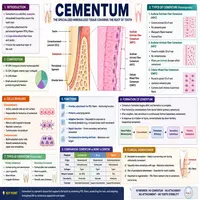
This root-covering tissue is made mostly of a mineralized matrix, along with collagen fibers and specialized cells. Its composition allows it to bond tightly with the periodontal ligament while still resisting resorption under normal chewing pressure. The table below outlines its main types and characteristics.
| Type | Location | Key Feature |
|---|---|---|
| Acellular cementum | Coronal two-thirds of the root | Forms before eruption; contains no embedded cells |
| Cellular cementum | Apical third and furcation areas | Forms after eruption; contains cementocytes |
| Afibrillar cementum | Cervical enamel margin | Lacks collagen fibers; thin protective layer |
| Cellular mixed stratified cementum | Apical region of multi-rooted teeth | Combines cellular and fibrous layers for repair |
Roughly 45 to 50 percent of this tissue consists of inorganic hydroxyapatite, while the remainder includes organic collagen and water. Consequently, it shares chemical similarities with bone, even though it functions quite differently since it has no internal blood vessels or nerves.
Functions of Cementum
This tissue performs several jobs that directly support tooth stability and periodontal health. Its primary roles generally fall into four categories.
- Attachment: It anchors Sharpey’s fibers from the periodontal ligament, securing the tooth within its socket.
- Protection: It covers and seals the dentin of the root, shielding it from bacterial invasion and external irritants.
- Compensation: It gradually thickens near the apex, helping maintain proper tooth length as occlusal surfaces wear down.
- Repair: It can form new layers over damaged root areas, sealing minor fractures or resorption defects.
Because this layer lacks its own blood supply, healing after injury tends to be slower than in vascularized tissues. Therefore, dentists monitor root surfaces carefully after trauma, since delayed repair can leave the area vulnerable to further breakdown.
How Cementum Forms: A Simple Flowchart
Formation of this tissue follows a defined biological sequence, beginning before a tooth even erupts into the mouth. The flowchart below outlines this process in simplified steps.
Hertwig's Epithelial Root Sheath Forms Around Developing Root
        │
        ▼
Root Sheath Breaks Apart, Exposing Underlying Dentin Surface
        │
        ▼
Cementoblasts Differentiate and Migrate to the Root Surface
        │
        ▼
Cementoblasts Deposit Collagen-Rich Matrix Onto the Root
        │
        ▼
Matrix Mineralizes, Forming a Hard Cementum Layer
        │
        ▼
Periodontal Ligament Fibers Embed Into the New Layer
        │
        ▼
Tooth Root Becomes Anchored Within the Alveolar Socket
This sequence explains why root development and periodontal attachment happen almost simultaneously. If any step is disrupted, such as through genetic conditions or early trauma, the tooth may erupt with weakened root attachment or abnormal root shape.
Common Problems Affecting Cementum
Several conditions can damage or alter this tissue, often with consequences for tooth stability and longevity.
| Condition | Cause | Effect |
|---|---|---|
| Cemental resorption | Trauma, orthodontic force, or chronic inflammation | Gradual loss of root surface tissue |
| Hypercementosis | Chronic inflammation or excessive occlusal force | Abnormal thickening of the root surface |
| Cementicles | Calcified nodules within the periodontal ligament | May interfere with normal ligament function |
| Exposed cementum | Gum recession from periodontal disease | Increased root sensitivity and decay risk |
Since this layer is softer than enamel, an exposed root surface decays more easily once gum recession occurs. As a result, patients with visible root surfaces often need targeted preventive care, including desensitizing treatments and careful hygiene instruction.
Concept Map: Cementum’s Role in Periodontal Health
To visualize how this tissue connects to broader dental function, consider the concept map below.
                          CEMENTUM
                             │
      ┌──────────────────────┼──────────────────────┐
      │                      │                        │
  STRUCTURE               FUNCTION                PROBLEMS
      │                      │                        │
 Acellular layer        Fiber attachment        Resorption
 Cellular layer         Root protection       Hypercementosis
 Hydroxyapatite         Wear compensation         Cementicles
 Collagen matrix         Repair capacity      Exposed root surface
      │                      │                        │
      └──────────────────────┼──────────────────────┘
                             │
                    TOOTH STABILITY & ROOT HEALTH
This map shows why periodontists rarely evaluate this tissue in isolation. When it weakens or becomes exposed, the effects typically extend into ligament attachment, root sensitivity, and overall periodontal stability.
Protecting Cementum for Long-Term Dental Health
Preventing damage to this root surface starts with controlling the factors that lead to gum recession and root exposure. Consistent brushing with a soft-bristled brush, along with gentle flossing, helps prevent the plaque buildup that drives periodontal disease. Meanwhile, routine dental visits allow early detection of recession before root surfaces become vulnerable.
Diet also plays a supporting role, since adequate calcium and vitamin D intake help maintain healthy mineralized tissues throughout the body, including the roots of teeth. On the other hand, aggressive brushing technique can accelerate gum recession, exposing root surfaces prematurely and increasing sensitivity.
Finally, patients undergoing orthodontic treatment should follow their provider’s guidance closely, since excessive or poorly controlled force can trigger resorption of this tissue. Regular monitoring during treatment helps catch early warning signs before permanent damage occurs.
Conclusion
Cementum may not be visible during a smile, but it plays an indispensable role in anchoring teeth securely within the jaw. It bridges the gap between root and bone, compensates for wear over a lifetime, and provides a repair mechanism when minor damage occurs. Understanding its structure and function clarifies why periodontists monitor root surfaces so carefully, particularly in patients with gum recession or a history of orthodontic treatment.
By maintaining consistent oral hygiene, eating a nutrient-rich diet, and following professional guidance during dental treatment, patients can protect this tissue for decades. Ultimately, healthy root coverage supports not just individual tooth stability, but the long-term strength of the entire periodontal system.
Frequently Asked Questions
It consists mainly of mineralized hydroxyapatite along with collagen fibers and water, giving it a composition similar to bone but without an internal blood supply.
Limited repair is possible, since cementoblasts can deposit new layers over minor defects. However, significant loss from resorption or trauma often requires professional periodontal intervention.
Once gum recession exposes the root surface, this softer tissue wears down more easily than enamel, allowing temperature and pressure changes to reach the nerve more directly.
Excessive or poorly controlled force during orthodontic movement can trigger resorption of the root surface. Careful force calibration by an orthodontist helps minimize this risk.
Use a soft-bristled toothbrush, avoid aggressive brushing technique, maintain a calcium-rich diet, attend regular dental checkups, and follow orthodontic guidance closely if undergoing treatment.