Periodontal Ligament: The Hidden Support System Behind Every Healthy Tooth
Every tooth in the mouth relies on a thin, fibrous layer that most people never think about. This tissue cushions chewing forces, anchors the tooth to the jawbone, and sends constant feedback to the brain about pressure and position. Dentists call it the periodontal ligament, and it plays a far bigger role in oral health than its small size suggests.
In this article, we break down what this tissue is, how it is built, what it does, and why keeping it healthy matters for long-term dental stability. Along the way, you will find tables, a simple flowchart, and a concept map to make the anatomy easier to visualize. By the end, you will understand why periodontists pay such close attention to this often-overlooked structure.
What Is the Periodontal Ligament?
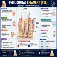
The periodontal ligament is a thin band of connective tissue that sits between the root of a tooth and the surrounding bone socket, known as the alveolar bone. Rather than fusing the tooth directly to bone, this ligament creates a flexible, shock-absorbing connection. Consequently, teeth can withstand the daily forces of biting and chewing without transferring damaging stress directly to the jaw.
This tissue measures only about 0.15 to 0.38 millimeters in width, yet it performs several critical jobs at once. It holds the tooth in place, nourishes surrounding structures, and acts as a sensory organ that detects pressure changes during chewing. Because it sits at the junction of tooth and bone, dentists often examine its condition when diagnosing gum disease or planning orthodontic treatment.
Interestingly, this ligament is not a static structure. It constantly remodels itself throughout life, adapting to the forces placed on teeth. As a result, orthodontic tooth movement is possible: braces and aligners work specifically because this tissue can reshape itself over time.
Anatomical Structure and Composition
The periodontal ligament is made up of collagen fibers, cells, blood vessels, and nerve endings, all working together within a soft connective tissue matrix. These components allow the ligament to be both strong and adaptable. Below is a breakdown of its main fiber groups and their roles.
| Fiber Group | Location | Primary Function |
|---|---|---|
| Alveolar crest fibers | Near the top of the socket | Resist tilting and lateral forces |
| Horizontal fibers | Below the crest fibers | Stabilize the tooth against sideways pressure |
| Oblique fibers | Largest group, mid-root region | Absorb vertical chewing forces |
| Apical fibers | Around the root tip | Prevent the tooth from being pulled out |
| Interradicular fibers | Between roots of multi-rooted teeth | Resist rotation and tipping |
In addition to collagen fibers, the ligament contains fibroblasts, cementoblasts, osteoblasts, and epithelial cell rests of Malassez. Fibroblasts continuously produce and break down collagen, therefore keeping the tissue in a state of ongoing renewal. Meanwhile, blood vessels supply nutrients, and nerve fibers provide the sensory feedback needed for proprioception, which is the ability to sense tooth position and pressure without looking.
Functions of the Periodontal Ligament
This connective tissue does far more than hold a tooth in its socket. Its functions generally fall into four categories, each essential to healthy oral function.
- Support and attachment: It suspends the tooth within the bone socket, distributing biting forces evenly.
- Shock absorption: It cushions the tooth root from sudden or repeated mechanical stress during chewing.
- Sensory feedback: Nerve endings within the ligament detect pressure, allowing the brain to adjust bite force instantly.
- Nutritive and remodeling function: Blood vessels nourish the tooth’s cementum, while cells continuously rebuild bone and ligament fibers.
Because of these combined roles, damage to the periodontal ligament can affect chewing comfort, tooth stability, and even speech in severe cases. Furthermore, since this tissue links directly to the bone, chronic inflammation here often signals the early stages of periodontal disease.
How the Periodontal Ligament Enables Tooth Movement
Orthodontic treatment depends entirely on the biological responses of this tissue. The process follows a predictable cycle, illustrated in the simplified flowchart below.
Force Applied to Tooth (e.g., braces)
        │
        ▼
Ligament Compresses on One Side, Stretches on the Other
        │
        ▼
Compressed Side: Bone Resorption Begins (Osteoclast Activity)
Stretched Side: New Bone Formation Begins (Osteoblast Activity)
        │
        ▼
Tooth Gradually Shifts Toward the Direction of Force
        │
        ▼
Ligament Fibers Reorganize Around the New Tooth Position
        │
        ▼
Tooth Stabilizes in Its New Location
This cycle explains why orthodontic adjustments happen gradually rather than all at once. Applying excessive force too quickly can damage the ligament or cause root resorption. Therefore, orthodontists carefully calibrate pressure to work with the body’s natural remodeling pace rather than against it.
Common Disorders Affecting the Periodontal Ligament
Several conditions can compromise the integrity of this ligament, often leading to tooth mobility or loss if left untreated.
| Condition | Cause | Effect on the Ligament |
|---|---|---|
| Periodontitis | Bacterial plaque and chronic inflammation | Breaks down collagen fibers and supporting bone |
| Traumatic occlusion | Excessive or uneven bite force | Causes widening and inflammation of the ligament space |
| Ankylosis | Fusion of tooth root directly to bone | Ligament space disappears, tooth loses mobility |
| Root resorption | Prolonged orthodontic force or trauma | Damages ligament attachment and root surface |
| Periodontal abscess | Localized bacterial infection | Rapid destruction of ligament and surrounding tissue |
Early detection matters greatly here. Dentists often use X-rays to check the width and continuity of the ligament space, since a widened or irregular space frequently points to underlying disease. Additionally, bleeding, swelling, or tooth mobility can serve as warning signs that this tissue is under stress.
Concept Map: How the Periodontal Ligament Connects to Oral Health
To visualize how this single tissue links to broader dental health, consider the following concept map.
                     PERIODONTAL LIGAMENT
                            │
      ┌─────────────────────┼─────────────────────┐
      │                     │                       │
  STRUCTURE              FUNCTION               DISORDERS
      │                     │                       │
 Collagen fibers      Shock absorption        Periodontitis
 Fibroblasts           Tooth support           Ankylosis
 Blood vessels        Sensory feedback       Root resorption
 Nerve endings        Bone remodeling        Traumatic occlusion
      │                     │                       │
      └─────────────────────┼─────────────────────┘
                            │
                    OVERALL ORAL HEALTH
                (chewing efficiency, tooth
                 stability, gum health)
This map highlights why periodontists treat the ligament as central to overall dental function rather than an isolated structure. When one branch weakens, the effects typically ripple outward into chewing efficiency, gum stability, and long-term tooth retention.
Maintaining a Healthy Periodontal Ligament
Protecting this tissue starts with consistent, practical habits rather than complicated routines. Brushing twice daily and flossing regularly remove the plaque that would otherwise trigger inflammation. Meanwhile, routine dental checkups allow professionals to catch early signs of ligament stress before symptoms become noticeable.
Diet also plays a meaningful role. Foods rich in vitamin C and calcium support collagen production and bone density, both of which strengthen the tissue surrounding each tooth. On the other hand, smoking significantly impairs blood flow to the gums, which slows healing and accelerates tissue breakdown.
Finally, avoiding excessive bite force, such as chronic teeth grinding, helps prevent unnecessary strain. Night guards, stress management, and orthodontic correction can all reduce this pressure and preserve the ligament’s structural integrity over time.
Conclusion
The periodontal ligament may be thin, but its impact on oral health is substantial. It anchors teeth firmly while allowing enough flexibility to absorb daily chewing forces, and it continuously remodels itself in response to pressure and movement. Understanding its structure and function helps explain why periodontists monitor it so closely during checkups, and why early intervention matters when signs of disease appear.
By maintaining good oral hygiene, eating a nutrient-rich diet, and addressing bite issues early, patients can protect this vital tissue for years to come. Ultimately, a healthy periodontal ligament supports not just individual teeth, but the long-term stability of the entire dental arch.
Frequently Asked Questions
It consists mainly of collagen fibers, along with fibroblasts, blood vessels, and nerve endings, all embedded in a soft connective tissue matrix between the tooth root and the alveolar bone.
Mild inflammation can often resolve with improved oral hygiene and professional cleaning. However, significant fiber damage or bone loss usually requires periodontal treatment to prevent further deterioration.
It allows teeth to move gradually by remodeling bone on either side of the root. Without this biological response, braces and aligners would not be able to reposition teeth safely.
Common signs include tooth mobility, gum swelling, bleeding while brushing, and sensitivity to pressure. A dentist can confirm the diagnosis using X-rays and a periodontal examination.
Brush and floss daily, schedule regular dental checkups, eat a balanced diet rich in vitamin C and calcium, avoid smoking, and manage teeth grinding with a night guard if needed.