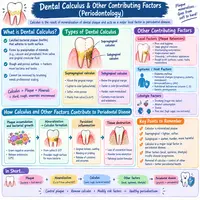
Dental Calculus and Other Contributing Factors in Gum Disease
Gum disease rarely comes from a single cause. Plaque starts the process, but other elements often push it further. Hardened deposits called dental calculus play a major role here. So do habits, health conditions, and even genetics. In this guide, we explore how calculus forms. We also cover other factors that contribute to gum disease. You will find two tables, a simple flowchart, and five common questions answered. Understanding these pieces helps you protect your gums more effectively.
What Is Dental Calculus?
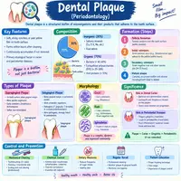
Calculus, often called tartar, is hardened plaque. Once soft plaque sits undisturbed, minerals from saliva begin depositing within it. Over time, this mineral buildup hardens into a rough, cement-like layer. Unlike plaque, calculus cannot be brushed away at home.
This hardened surface creates a rough texture on your teeth. That roughness actually attracts more plaque, creating a frustrating cycle. Fresh bacteria cling easily to its uneven surface. Consequently, calculus does not just sit passively; it actively worsens the problem.
Calculus forms both above and below the gumline. Supragingival calculus appears above the gum, often visible as a yellow or tan buildup. Subgingival calculus forms below the gumline, hidden from view, and often darker in color. Both types require professional removal, since no home tool can safely eliminate them.
How Calculus Forms: The Process Explained
Calculus formation follows a fairly predictable timeline. Plaque begins forming within minutes after brushing. Without removal, this soft layer matures within about a day. From there, mineralization begins if the plaque remains undisturbed.
Saliva carries calcium and phosphate minerals naturally. These minerals gradually deposit into the plaque’s sticky matrix. Within several days, the plaque can start hardening noticeably. After one to two weeks, this process often produces firmly attached calculus.
Certain areas mineralize faster than others. Saliva flows more heavily near the lower front teeth and upper back molars. As a result, calculus tends to build up fastest in these specific zones. Recognizing these high-risk areas matters. It helps explain why dentists focus extra attention there during exams.
How Dentists Detect Calculus
Spotting calculus takes a trained eye and the right tools. Supragingival deposits are often visible during a routine visual exam. A dental explorer, a thin metal probe, helps confirm rough or hardened areas. Your dentist gently runs it along the tooth surface.
Subgingival calculus requires more careful detection. Probing depths reveal pockets where hidden deposits may lurk. X-rays sometimes show larger calculus deposits as faint shadows near the root. Still, much subgingival calculus remains invisible even on imaging.
Because of this, regular professional exams matter enormously. Hygienists use tactile feel, not just sight, to detect rough subgingival surfaces. This hands-on skill catches buildup that imaging might miss for months or years.
Why Calculus Is So Harmful to Your Gums
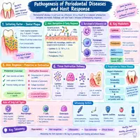
Calculus does more than simply look unpleasant. Its rough surface irritates the surrounding gum tissue constantly. This ongoing irritation triggers chronic inflammation along the gumline. Over time, that inflammation can progress toward more serious gum disease.
Subgingival calculus poses an even greater risk. Hidden below the gumline, it sits directly against vulnerable tissue. Bacteria cling to its surface, releasing toxins that damage the gum and bone. Patients cannot easily see or feel this buildup. As a result, it often goes unnoticed for a long time.
Removing calculus becomes essential for gum health, not optional. Scaling procedures use specialized tools to break the hardened deposits free. Professional cleanings reach areas that daily brushing simply cannot access. Regular removal interrupts the cycle of irritation and prevents further tissue damage.
Local Contributing Factors Beyond Calculus
Calculus is not the only local factor at play. Several other elements in the mouth can worsen gum disease. Overhanging or poorly fitted dental restorations trap plaque against the gum. Crowded or crooked teeth also create hard-to-clean spaces.
Mouth breathing dries out gum tissue, making it more vulnerable to irritation. Food impaction between teeth creates another persistent irritant. Orthodontic appliances, while helpful long term, can trap plaque around brackets and wires. Each of these factors adds its own layer of risk.
Habits matter too. Grinding or clenching places extra stress on gum and bone tissue. Poor-fitting partial dentures can irritate nearby gum areas repeatedly. The table below summarizes common local factors that worsen gum disease beyond calculus alone.
| Local Factor | How It Contributes |
|---|---|
| Overhanging restorations | Trap plaque against the gum edge |
| Crowded or crooked teeth | Create hard-to-clean spaces |
| Mouth breathing | Dries and irritates gum tissue |
| Orthodontic appliances | Trap plaque around brackets and wires |
| Food impaction | Causes persistent local irritation |
| Teeth grinding | Adds mechanical stress to supporting tissue |
Systemic Factors That Worsen Gum Disease
Local factors tell only part of the story. Several whole-body conditions influence gum health significantly. Diabetes stands out as one of the strongest contributors. Poorly controlled blood sugar fuels inflammation and slows healing throughout the body.
Hormonal changes also play a notable role. Pregnancy, puberty, and menopause can all heighten gum sensitivity to plaque and calculus. Certain medications, including some blood pressure drugs and anticonvulsants, can cause gum overgrowth. This overgrowth creates additional spaces where plaque and calculus accumulate.
Smoking deserves special mention here too. It reduces blood flow to the gums, masking visible symptoms like bleeding. Ironically, this can delay diagnosis while damage continues underneath. Genetics and immune function round out this picture. Some people simply react more strongly to the same bacterial load.
The Role of Diet and Nutrition
What you eat shapes your calculus risk too. Diets high in refined sugar feed plaque-forming bacteria rapidly. This accelerates the whole mineralization process described earlier. Reducing sugary snacks and drinks slows plaque buildup meaningfully.
Certain nutrients support healthy gum tissue directly. Vitamin C plays a key role in collagen production and gum repair. Calcium and vitamin D support the bone that holds your teeth in place. A balanced diet strengthens your defenses against inflammation overall.
Hydration matters as well. Saliva naturally rinses away food particles and buffers harmful acids. Chronic dry mouth, from dehydration or certain medications, raises calculus and decay risk. Drinking enough water throughout the day supports this natural defense system.
How These Factors Interact: A Simple Flowchart
These contributing factors rarely act alone. The flowchart below shows how they typically combine to worsen gum disease.
          Plaque forms on tooth surfaces
                        |
                        v
      Without removal, plaque hardens into calculus
                        |
                        v
   Local factors add further irritation
   (crowding, restorations, food impaction)
                        |
                        v
   Systemic factors amplify the body's response
   (diabetes, hormones, smoking, genetics)
                        |
                        v
      Chronic gum inflammation develops
                        |
                        v
   Without treatment, periodontitis may progress
                        |
                        v
   Professional cleaning and risk management
        interrupt this cycle
Managing These Risk Factors Effectively
Addressing contributing factors requires a layered approach. Professional cleanings remain essential for removing hardened calculus safely. Most dentists recommend cleanings every six months. Higher-risk patients may need more frequent visits instead.
Daily home care still matters enormously. Thorough brushing and flossing slow new plaque formation between visits. Addressing local irritants also helps significantly. Replacing an overhanging filling helps significantly. So does adjusting an ill-fitting denture that irritates the gum.
Managing systemic health pays real dividends too. Keeping blood sugar well controlled reduces inflammation throughout the body. Quitting smoking improves blood flow and healing capacity substantially. Working closely with your dentist and physician helps most. Together, they offer complete protection against gum disease.
Conclusion
Dental calculus rarely acts alone in driving gum disease forward. As we have seen, local irritants and systemic health often work together. Hardened plaque adds its own layer too. Crowded teeth, smoking, diabetes, and hormonal shifts all add their own layer of risk. Fortunately, understanding these contributing factors empowers better prevention. Regular professional cleanings remain your strongest defense. Consistent home care and attention to overall health help too. Addressing every piece of this puzzle protects your gums for the long run.
Frequently Asked Questions
No. Calculus hardens onto the tooth surface and requires professional scaling tools for safe removal.
Saliva flow concentrates near the lower front teeth and upper molars. This higher mineral exposure speeds up calculus formation in those areas.
Yes. Smoking reduces blood flow to the gums. This masks symptoms like bleeding, while damage continues underneath.
Most patients benefit from cleanings every six months. Higher-risk patients, including smokers or those with diabetes, may need more frequent visits.
Yes. Crowded or crooked teeth create spaces that are harder to clean. This allows plaque and calculus to build up more easily.