Cell Injury: Causes, Types, and Mechanisms
Every disease process starts somewhere at the cellular level. Something disrupts a cell’s normal balance, and the cell responds in predictable ways. Pathologists study this response closely because it explains almost every illness humans experience. From heart attacks to liver disease, the same underlying biology repeats itself again and again. This guide walks through the causes, stages, and clinical relevance of this core pathology concept in plain, practical language. By the end, you’ll understand exactly why a damaged cell either bounces back or dies for good.
Understanding the Basics
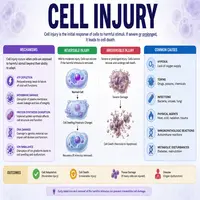
Cell injury happens when a cell can no longer maintain its normal internal environment. Stress factors overwhelm the cell’s adaptive capacity, and structural or functional damage follows. Initially, cells try to adapt through mechanisms like hypertrophy or atrophy. However, once the stress exceeds the cell’s tolerance, true injury begins.
Two outcomes are possible after this point. First, the damage may stay reversible, meaning the cell recovers once the stressor disappears. Second, the damage may become irreversible, pushing the cell toward death. Understanding where that tipping point sits remains central to modern pathology.
Common Causes Behind Cellular Damage
Multiple factors can trigger this process, and most fall into recognizable categories. Oxygen deprivation, known as hypoxia, ranks among the most frequent causes. Blocked blood flow, severe anemia, or lung disease can all starve tissue of oxygen. Consequently, mitochondria fail first because they depend heavily on oxygen for energy production.
Physical agents also play a major role. Trauma, extreme temperatures, and radiation exposure damage cell membranes directly. Meanwhile, chemical agents such as toxins, drugs, and pollutants interfere with enzyme function and membrane integrity. Infectious organisms, including bacteria, viruses, and parasites, contribute significantly too.
Nutritional imbalances deserve mention as well. Both excess and deficiency states harm cells over time. For instance, vitamin deficiencies impair enzyme activity, while excess fat accumulation stresses the liver. Genetic mutations round out the list, since inherited defects can weaken a cell’s structure from birth. Immunologic reactions add yet another layer, as the immune system sometimes attacks the body’s own healthy tissue by mistake.
Adaptive Responses Before True Damage Sets In
Cells rarely surrender to stress immediately. Instead, they attempt several adaptive strategies first. Hypertrophy allows individual cells to grow larger, which explains why heart muscle thickens under chronic high blood pressure. Hyperplasia, by contrast, increases cell number rather than size, as seen in the uterus during pregnancy.
Atrophy works in the opposite direction, shrinking cells that receive less use or blood supply. Metaplasia, meanwhile, swaps one mature cell type for another better suited to handle ongoing stress. Smokers’ airways, for example, often replace normal ciliated cells with tougher squamous cells over time.
These adaptations buy the cell valuable time. However, once the stimulus becomes too intense or prolonged, adaptation fails and true damage begins.
Reversible vs Irreversible Damage
Distinguishing these two categories matters enormously in clinical practice. The table below highlights the key differences.
| Feature | Reversible Damage | Irreversible Damage |
|---|---|---|
| Cell membrane | Intact | Severely disrupted |
| Mitochondria | Swollen but functional | Permanently damaged |
| Nucleus | Normal | Pyknosis, karyorrhexis, karyolysis |
| Outcome | Full recovery possible | Cell death occurs |
| Example | Cellular swelling | Coagulative necrosis |
Reversible changes typically show up as cellular swelling and fatty change. Cells appear larger under the microscope, and organelles look distorted but salvageable. Removing the harmful stimulus at this stage restores normal function.
Irreversible changes, on the other hand, cross a point of no return. Membrane damage becomes severe, calcium floods into the cell, and enzymes begin digesting cellular components. Eventually, the nucleus breaks down entirely, confirming that death has occurred.
A Simple Flowchart: How the Process Unfolds
Below is a quick visual pathway showing the typical progression.
Harmful stimulus applied
        |
        v
Cell attempts adaptation
        |
        v
   Stress within tolerance?  ---Yes---> Cell adapts (hypertrophy, atrophy)
        |No
        v
Cellular stress response triggered
        |
        v
   Damage reversible?  ---Yes---> Stimulus removed --> Full recovery
        |No
        v
Membrane and mitochondrial failure
        |
        v
Calcium influx and enzyme activation
        |
        v
Necrosis or apoptosis occurs
This pathway explains why early intervention matters so much in medicine. Once the process crosses into the irreversible zone, no treatment can reverse the outcome.
Morphological Changes Seen Under the Microscope
Pathologists rely on specific visual clues to identify damaged tissue. Cellular swelling appears first, caused by failure of the sodium-potassium pump. Water and sodium accumulate inside the cell, making it look puffy and pale.
Fatty change follows in metabolically active organs like the liver. Lipid droplets accumulate within the cytoplasm, disrupting normal architecture. Subsequently, if the stress continues, structural proteins denature and membranes rupture.
Nuclear changes offer the clearest sign of a point of no return. Pyknosis causes the nucleus to shrink and darken. Karyorrhexis follows, fragmenting the nucleus into small pieces. Finally, karyolysis dissolves the nucleus completely, leaving no trace behind.
Types of Necrosis Worth Knowing
Necrosis doesn’t look the same everywhere in the body. Different tissues die in distinct patterns, and pathologists use these patterns to identify the underlying cause.
| Necrosis Type | Typical Location | Common Cause |
|---|---|---|
| Coagulative | Heart, kidney | Ischemia (loss of blood flow) |
| Liquefactive | Brain | Ischemia, bacterial infection |
| Caseous | Lungs (tuberculosis) | Mycobacterial infection |
| Fat necrosis | Pancreas, breast tissue | Enzymatic digestion, trauma |
| Gangrenous | Limbs, GI tract | Prolonged ischemia |
Coagulative necrosis preserves the tissue’s general architecture for several days, which helps pathologists date the injury. Liquefactive necrosis, on the other hand, dissolves tissue into a liquid mass almost immediately. Caseous necrosis produces a cheese-like, crumbly appearance under the microscope, while fat necrosis creates chalky white deposits from released fatty acids.
Clinical Significance and Real-World Relevance
Doctors apply this knowledge across nearly every specialty. Cardiologists track ischemic damage after a heart attack, since dead tissue cannot pump blood effectively. Neurologists monitor stroke patients closely because brain cells tolerate oxygen loss poorly. Nephrologists watch for acute tubular necrosis following prolonged low blood pressure.
Surgeons also consider this concept before major operations. Prolonged clamping of blood vessels risks starving downstream tissue of oxygen. Therefore, surgical teams carefully time each step to minimize damage. Pharmacologists, meanwhile, study how certain drugs protect cells during periods of stress, opening doors to new treatment strategies.
Furthermore, researchers continue exploring how chronic, low-grade damage contributes to aging and degenerative disease. Emerging evidence links repeated cellular stress to conditions like Alzheimer’s disease and chronic kidney disease. As a result, this field keeps expanding well beyond its traditional boundaries in modern medicine.
Conclusion
Cell injury sits at the foundation of nearly every disease process doctors encounter. Recognizing the difference between reversible and irreversible damage helps clinicians decide how aggressively to intervene. Early detection often means the difference between full recovery and permanent tissue loss. As research uncovers new protective mechanisms, treatment options for hypoxic and toxic damage continue improving. Ultimately, mastering this concept gives healthcare professionals a sharper lens for understanding disease at its earliest, most treatable stage.
Frequently Asked Questions
Oxygen deprivation, known as hypoxia, ranks as the most frequent trigger, followed closely by physical trauma and toxic chemical exposure.
Reversible changes show intact membranes and functional mitochondria under the microscope, while irreversible changes show membrane rupture and nuclear breakdown.
Necrosis is uncontrolled cell death caused by external injury, while apoptosis is a programmed, controlled process the cell triggers internally.
No, recovery depends on the severity and duration of the stressor. Once mitochondrial and membrane damage becomes severe, the process becomes irreversible
It explains the biological basis behind heart attacks, strokes, kidney failure, and many chronic diseases, guiding how doctors diagnose and treat patients.