Gingiva Explained: Anatomy, Health, and Common Disorders
Every periodontal exam begins with a close look at the soft tissue surrounding the teeth, yet many patients and even dental students overlook how complex this structure really is. However, understanding its anatomy and function helps explain why gum health influences the entire oral cavity. Consequently, learning about the gingiva in detail becomes essential before studying periodontal disease or planning any restorative treatment. In this article, we will explore its anatomy, examine common disorders, and outline diagnostic and preventive strategies. Additionally, we will use tables, a flowchart, and a concept map to simplify the material. Therefore, whether you are a student or a curious patient, this guide should deepen your understanding of a tissue that plays a much bigger role than most people realize.
What Is This Tissue?
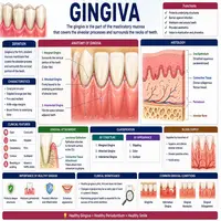
This soft tissue forms the visible pink collar surrounding each tooth and covers the alveolar bone beneath it. Structurally, it consists of epithelium and underlying connective tissue that together create a protective barrier against bacteria and mechanical irritation. Moreover, healthy tissue typically appears coral pink, firm, and stippled, resembling the texture of an orange peel. As a result, any deviation from this appearance, such as swelling, redness, or bleeding, often signals early inflammation. Furthermore, this tissue attaches tightly to the tooth surface through a specialized junctional epithelium, which forms a seal that prevents bacteria from reaching deeper structures.
Nevertheless, this seal can weaken when plaque accumulates along the gumline. Once bacteria penetrate this protective barrier, inflammation frequently develops, setting the stage for more serious periodontal problems if left unaddressed. Therefore, dentists closely monitor this junction during every routine examination.
Anatomical Zones
Clinicians typically divide this tissue into distinct anatomical zones, each serving a specific protective function. The table below summarizes these regions along with their key characteristics.
| Zone | Location | Key Feature |
|---|---|---|
| Free Margin | Unattached edge nearest the tooth | Forms the gingival sulcus |
| Attached Zone | Firmly bound to underlying bone | Provides stability during chewing |
| Interdental Papilla | Fills the space between adjacent teeth | Prevents food impaction |
| Mucogingival Junction | Border between attached tissue and alveolar mucosa | Marks transition to looser tissue |
Generally, each zone contributes to overall stability and protection against bacterial invasion. On the other hand, disruption in any one area, such as recession at the free margin, can expose sensitive root surfaces and increase disease risk.
Blood supply and nerve distribution also shape how this tissue responds to injury and infection. Small vessels branch from the alveolar arteries, delivering oxygen and nutrients that support rapid healing after minor trauma. Similarly, sensory nerve fibers make this tissue quite responsive to temperature changes and mechanical pressure, which explains why inflammation often causes noticeable discomfort. When plaque irritates the free margin over time, these vessels dilate, producing the redness and swelling characteristic of early gingivitis. Therefore, understanding this vascular and neural relationship helps clinicians interpret clinical signs more accurately during routine examinations.
From Health to Disease: A Flowchart
Bacterial plaque accumulation typically follows a predictable pattern before progressing into more advanced periodontal disease. The flowchart below illustrates this pathway step by step.
Flowchart: Progression of Periodontal Disease
Step 1: Plaque accumulates along the gumline
        |
        v
Step 2: Bacterial toxins irritate the tissue
        |
        v
Step 3: Inflammation develops (gingivitis)
        |
        v
Step 4: Bleeding and swelling become noticeable
        |
        v
Step 5: Untreated inflammation spreads to supporting bone
        |
        v
Step 6: Periodontal ligament and bone begin breaking down
        |
        v
Step 7: Periodontitis develops, risking tooth loss
This progression highlights why early intervention matters so much. Furthermore, gingivitis remains fully reversible with proper oral hygiene and professional cleaning. Eventually, if inflammation persists untreated, the damage becomes permanent once bone loss begins, making prevention far more valuable than treatment at later stages.
Common Disorders
Several conditions can affect this tissue, ranging from mild irritation to severe disease. Gingivitis, the most common disorder, causes redness, swelling, and bleeding due to plaque-induced inflammation. In contrast, periodontitis represents a more advanced stage where inflammation has damaged the supporting bone and ligament. Similarly, gingival recession occurs when tissue pulls away from the tooth, often exposing sensitive root surfaces. Meanwhile, gingival hyperplasia describes tissue overgrowth, sometimes triggered by certain medications, hormonal changes, or poor plaque control. In addition, acute necrotizing conditions, though less common, cause painful ulceration and require prompt professional treatment. Therefore, recognizing these distinct patterns helps clinicians select the most appropriate management strategy for each patient.
Concept Map: Disease Classification
To visualize how these conditions relate to one another, the table below organizes them under a shared structural theme.
| Level | Category | Sub-Category / Example |
|---|---|---|
| 1 | Periodontal Soft Tissue Health | Umbrella term |
| 2 | Inflammatory Conditions | Gingivitis, Periodontitis |
| 2 | Structural Changes | Recession, Hyperplasia |
| 3 | Clinical Response | Scaling, Root Planing, Surgical Therapy |
This layered structure shows how untreated inflammation can eventually progress into structural damage requiring more advanced intervention. Consequently, clinicians rarely view these conditions as isolated, unrelated events.
Diagnostic Approaches
Accurate diagnosis relies on a thorough clinical examination combined with periodontal probing. Dentists routinely measure pocket depth, assess bleeding on probing, and evaluate attachment loss to determine disease severity. Additionally, radiographs help reveal bone loss patterns that clinical examination alone cannot detect. Sometimes, clinicians also assess tissue color, texture, and contour, since subtle changes often indicate early inflammation before more obvious symptoms appear. As a result, combining multiple diagnostic methods significantly improves accuracy and allows earlier, more effective intervention.
Patient history also plays an important diagnostic role. For instance, smoking status, diabetes control, and hormonal changes all influence how tissue responds to bacterial irritation. Similarly, certain medications, such as calcium channel blockers, can trigger tissue overgrowth independent of plaque levels. Meanwhile, genetic predisposition may explain why some patients develop severe disease despite good oral hygiene. Consequently, a comprehensive evaluation considers both local and systemic factors before finalizing a diagnosis.
Prevention and Maintenance
Preventing disease in this tissue remains far easier than reversing advanced damage. Regular brushing and flossing disrupt plaque before it can trigger inflammation, while routine professional cleanings remove hardened deposits that home care cannot reach. In addition, patients who avoid tobacco use and maintain balanced blood sugar levels significantly reduce their risk of periodontal complications. Afterward, periodic periodontal maintenance visits help detect early changes before they progress into irreversible bone loss. Ultimately, consistent preventive care protects both the supporting tissue and the long-term stability of the teeth it surrounds.
Diet also contributes meaningfully to tissue resilience. Foods rich in vitamin C and antioxidants support collagen repair and help the body fight low-grade inflammation more effectively. In contrast, diets high in refined sugar feed plaque-forming bacteria, accelerating the inflammatory process described earlier. Similarly, adequate hydration supports healthy saliva flow, which naturally helps clear food debris and buffer acids throughout the day. Patients who combine good nutrition with mechanical plaque control typically experience fewer flare-ups and faster recovery when minor irritation does occur.
Conclusion
The gingiva serves as far more than a simple pink border around the teeth; it acts as the body’s first line of defense against oral bacteria. From its layered anatomy to the predictable progression of inflammatory disease, understanding this tissue equips both clinicians and patients to recognize problems early. Although gingivitis remains reversible with timely care, untreated inflammation can progress into permanent bone and attachment loss. Ultimately, consistent oral hygiene, professional monitoring, and prompt treatment together preserve this vital tissue and support long-term periodontal health.
Frequently Asked Questions
Healthy tissue typically appears coral pink, firm, and slightly stippled, without any swelling, redness, or bleeding during brushing.
Yes, gingivitis is fully reversible with consistent oral hygiene and professional cleaning, provided treatment begins before bone loss occurs
Recession often results from aggressive brushing, periodontal disease, or genetic thinness of the tissue, exposing sensitive root surfaces over time.
Most patients benefit from a professional evaluation every six months, though those with periodontal disease may need more frequent monitoring.
Yes, certain drugs, including calcium channel blockers and some anti-seizure medications, can cause tissue overgrowth independent of plaque control.