Spinal Cord Anatomy: Structure, Function, and Key Facts
The human body relies on one remarkable structure to carry messages between the brain and every muscle, organ, and limb. This structure is the spinal cord. It acts as the main communication highway of the nervous system. Without it, the brain could not control movement, sense touch, or manage vital reflexes.
In this guide, we explore the anatomy, layers, segments, and functions of this vital structure. We also cover common disorders, protective mechanisms, and frequently asked questions. Whether you are a student, healthcare professional, or curious reader, this article breaks down complex anatomy into simple, digestible sections.
What Is the Spinal Cord?
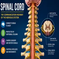
The spinal cord is a long, tube-shaped bundle of nerve tissue. It extends from the base of the brain down through the vertebral column. Specifically, it runs from the medulla oblongata to the first or second lumbar vertebra in adults.
This structure measures roughly 45 centimeters in length and about 1 centimeter in diameter. Despite its slim size, it carries millions of nerve fibers. These fibers transmit signals in both directions, connecting the brain to the rest of the body.
Additionally, this vital pathway is protected by three layers of connective tissue called meninges. These layers include the dura mater, arachnoid mater, and pia mater. Together, they cushion the delicate tissue and prevent injury from everyday movement.
Structure and Segments

The spinal cord is divided into four major regions. Each region corresponds to a specific area of the vertebral column and controls particular body functions.
| Region | Vertebrae Level | Number of Segments | Main Function |
|---|---|---|---|
| Cervical | C1–C7 | 8 | Neck, arms, diaphragm control |
| Thoracic | T1–T12 | 12 | Chest, abdominal muscles |
| Lumbar | L1–L5 | 5 | Hip and leg movement |
| Sacral | S1–S5 | 5 | Bladder, bowel, sexual function |
Furthermore, each segment gives rise to a pair of spinal nerves. These nerves exit through small openings called intervertebral foramina. As a result, every segment supplies sensation and movement to a specific part of the body.
Consequently, damage at any level can affect everything below that point. For example, an injury in the cervical region often causes wider paralysis than one in the lumbar region.
Internal Anatomy: Gray and White Matter

Inside the spinal cord, two distinct types of tissue perform different jobs. Understanding this internal layout helps explain how signals travel so efficiently.
Gray matter sits at the center and forms a butterfly-shaped pattern. It contains nerve cell bodies, dendrites, and synapses. Moreover, it processes reflexes and connects sensory input to motor output.
White matter surrounds the gray matter. It consists mainly of myelinated axons, which transmit signals rapidly. These fibers organize into tracts. Some tracts carry information upward to the brain, while others send commands downward to muscles.
In short, this dual structure allows fast, organized communication throughout the body. Therefore, any damage to either type of tissue can disrupt movement, sensation, or both.
Concept Map: How Signals Travel
Below is a simplified flow of how a sensory signal reaches the brain and how a motor response returns to the muscle.
SENSORY PATHWAY (Ascending)
Skin/Muscle Receptor → Peripheral Nerve → Dorsal Root → Spinal Cord (White Matter Tracts) → Brainstem → Thalamus → Brain (Sensory Cortex)

MOTOR PATHWAY (Descending)
Brain (Motor Cortex) → Brainstem → Spinal Cord (White Matter Tracts) → Ventral Root → Peripheral Nerve → Muscle
This two-way flow shows why the spinal cord is often called a “relay station.” Similarly, it explains why reflexes, such as pulling a hand away from heat, happen before the brain even processes the pain.
Key Functions
The spinal cord performs three essential roles in the body. First, it relays sensory information from the skin, muscles, and organs to the brain. Second, it carries motor commands from the brain to muscles, enabling voluntary movement. Third, it controls many reflexes independently, without needing brain involvement.
For instance, the knee-jerk reflex occurs entirely within this structure. A doctor taps the tendon, and the leg kicks almost instantly. This happens because the reflex arc completes locally, bypassing the brain for speed.
Additionally, this structure regulates involuntary functions like bladder control and blood pressure adjustments. Therefore, it plays a role far beyond simple movement.
Protective Mechanisms
Given its importance, the body protects this structure through multiple layers. The vertebral column forms a bony shield around it. Meanwhile, cerebrospinal fluid cushions it against shock and sudden impact.
The meninges add another layer of defense. The dura mater is tough and outermost. The arachnoid mater sits in the middle, resembling a spider’s web. Finally, the pia mater clings directly to the tissue, supplying it with blood vessels.
Together, these protective layers reduce the risk of injury during daily activities like running, jumping, or bending. However, severe trauma can still overwhelm these defenses.
Common Disorders and Injuries
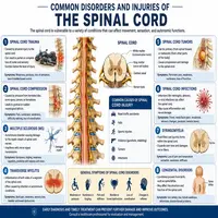
Several conditions can affect this critical structure. Below is a quick reference table.
| Condition | Cause | Common Symptoms |
|---|---|---|
| Spinal Cord Injury (SCI) | Trauma, accidents | Paralysis, loss of sensation |
| Multiple Sclerosis | Autoimmune demyelination | Weakness, numbness, vision issues |
| Spinal Stenosis | Narrowing of the canal | Pain, tingling, weakness |
| Transverse Myelitis | Inflammation | Sudden pain, motor loss |
| Syringomyelia | Fluid-filled cyst | Chronic pain, stiffness |
Early diagnosis often improves outcomes significantly. Consequently, doctors recommend prompt imaging, such as an MRI, whenever symptoms appear suddenly.
Why Spinal Cord Health Matters
Maintaining a healthy nervous system requires attention to posture, exercise, and injury prevention. Regular stretching keeps the vertebral column flexible. Meanwhile, core-strengthening exercises support proper alignment and reduce pressure on nerve tissue.
Equally important, wearing protective gear during sports lowers injury risk. Simple habits, like using seatbelts and avoiding risky dives into shallow water, prevent many traumatic injuries each year.
In addition, routine checkups help detect early signs of nerve compression or degeneration. Consequently, small issues get treated before they become severe.
Conclusion
The spinal cord stands as one of the most essential structures in human anatomy. It connects the brain to the body, controls reflexes, and manages both voluntary and involuntary functions. Its layered protection, from bone to fluid to membrane, reflects just how vital it is to survival and daily function.
Understanding its structure, segments, and pathways helps explain many aspects of human movement and sensation. Ultimately, protecting this structure through healthy habits and prompt medical care ensures long-term mobility and quality of life.
Frequently Asked Questions
It relays messages between the brain and body while also controlling many reflexes independently.
It measures approximately 45 centimeters in adults, extending from the brainstem to the lumbar region.
Unlike some tissues, nerve fibers here have limited natural repair ability, though ongoing research explores new treatments.
The vertebral column, cerebrospinal fluid, and three meningeal layers all work together for protection.
Damage can cause paralysis, loss of sensation, or bladder and bowel dysfunction, depending on the injury’s location and severity.